. 2007 Apr;15(4):499-509.
doi: 10.1016/j.str.2007.03.003.

Structural basis for selective inhibition of Mycobacterium tuberculosis protein tyrosine phosphatase PtpB

Affiliations

Structural basis for selective inhibition of Mycobacterium tuberculosis protein tyrosine phosphatase PtpB

Christoph Grundner et al. Structure. 2007 Apr.

Abstract

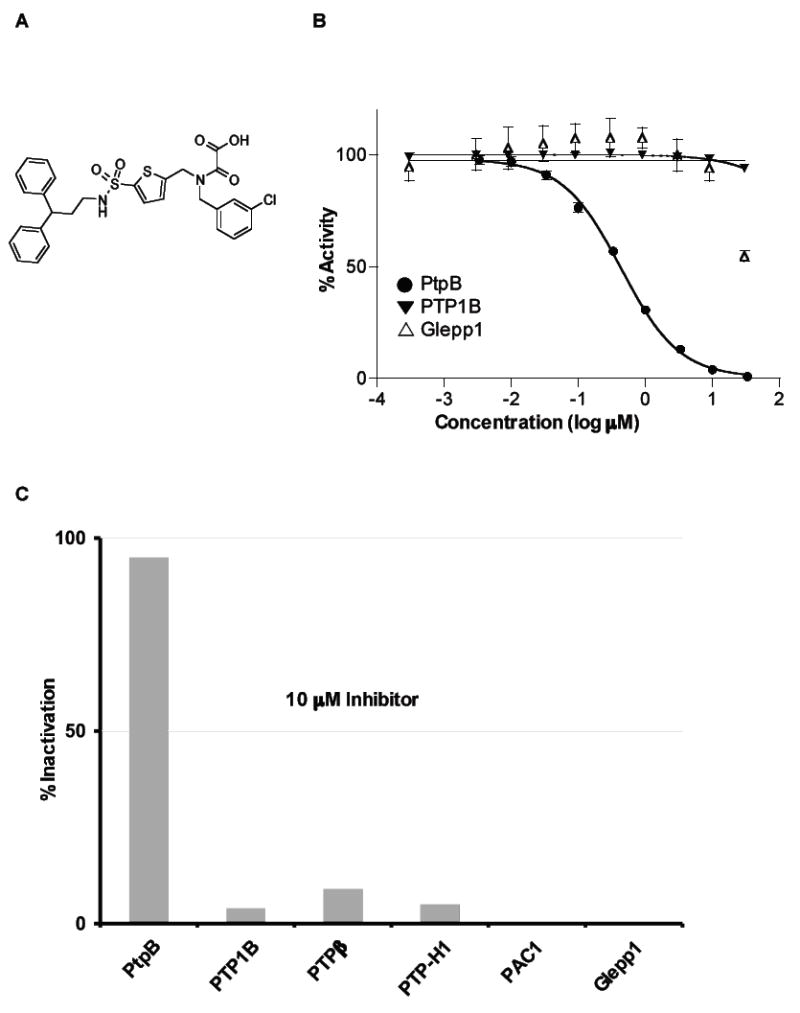
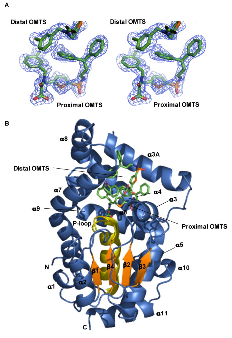
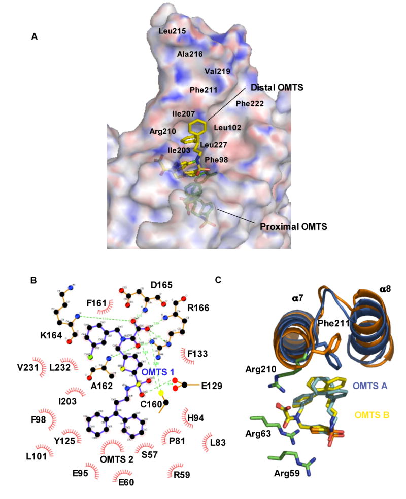
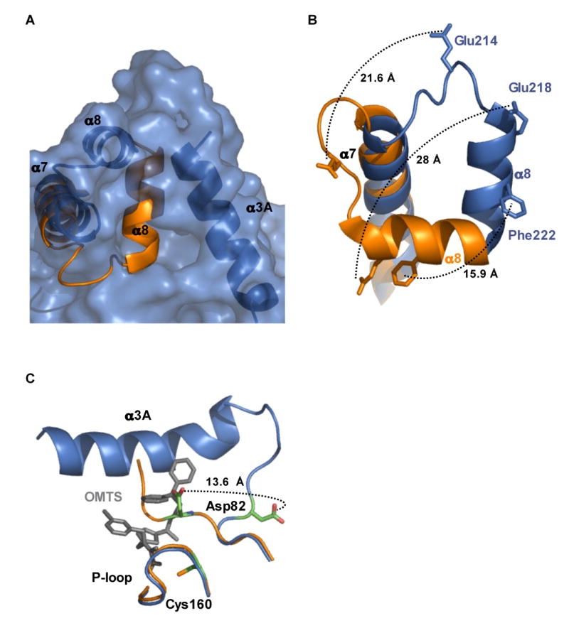
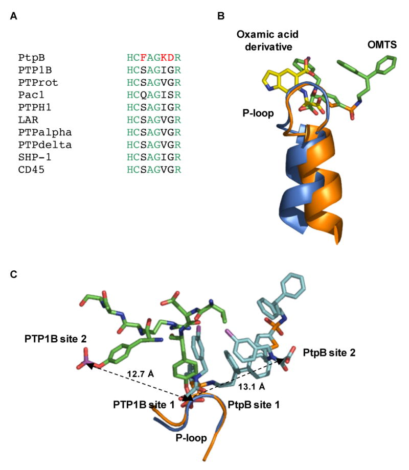
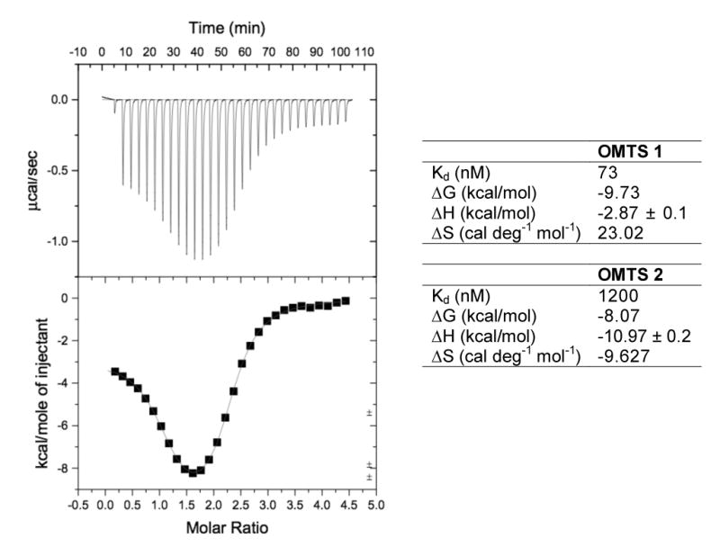
Tyrosine kinases and phosphatases establish the crucial balance of tyrosine phosphorylation in cellular signaling, but creating specific inhibitors of protein Tyr phosphatases (PTPs) remains a challenge. Here, we report the development of a potent, selective inhibitor of Mycobacterium tuberculosis PtpB, a bacterial PTP that is secreted into host cells where it disrupts unidentified signaling pathways. The inhibitor, (oxalylamino-methylene)-thiophene sulfonamide (OMTS), showed an IC(50) of 440 +/- 50 nM and >60-fold specificity for PtpB over six human PTPs. The 2 A resolution crystal structure of PtpB in complex with OMTS revealed a large rearrangement of the enzyme, with some residues shifting >27 A relative to the PtpB:PO(4) complex. Extensive contacts with the catalytic loop provide a potential basis for inhibitor selectivity. Two OMTS molecules bound adjacent to each other, raising the possibility of a second substrate phosphotyrosine binding site in PtpB. The PtpB:OMTS structure provides an unanticipated framework to guide inhibitor improvement.


Figures

Figure 1
Figure 1. Inhibitory activity of OMTS on phosphatases
(A) Structure of OMTS. (B) IC50 determination. Inhibitory activity of increasing concentrations of OMTS was assessed on PtpB, Glepp1 and PTP1B phosphatases. Mean values and standard deviations are represented. An IC50 value for PtpB of 0.44 μM was calculated from this experiment. (C) Selectivity of OMTS on a panel of phosphatases. Results are expressed as percent inhibition at 10 μM OMTS.
Figure 2
Figure 2. Inhibition kinetics
(A) Michaelis-Menten representation of PtpB inhibition. RFU: Relative fluorescence units. (B) Lineweaver-Burk representation of PtpB activity at increasing concentrations of inhibitor. Both representations indicate that OMTS is a competitive inhibitor of PtpB.
Figure 3
Figure 3. Overall structure of the PtpB:OMTS complex
(A) Stereo representation of the simulated-annealing, Fo-Fc omit map of OMTS bound in the PtpB active site contoured at 3 σ. Electron density for the proximal and the chlorobenzyl group of the distal OMTS is shown. (B) Two molecules of OMTS bind in the PtpB active site. Helix α8, part of the autoinhibitory lid, rotated around Ser228 by ∼90° to open the active site. Residues 88–103 form helix α3A not seen in the PtpB:PO4 structure. Helices α3, α3A, α4 and the α7–α8 hairpin ring the active-site cavity.
Figure 4
Figure 4. Interactions between OMTS and PtpB
(A) Electrostatic surface representation of PtpB generated with APBS (Baker et al., 2001. Blue: positive potential, red: negative potential) and stick representation of the inhibitors shows the depth of the active-site cleft. The distal OMTS molecule is more exposed, while the inhibitor proximal to Cys160 is buried. The approximate locations of hydrophobic residues lining the surface of the helix-α7–α8 hairpin are indicated. (B) Schematic drawing of interactions between the proximal inhibitor and the PtpB catalytic site (generated with LIGPLOT and edited manually). Water 391 contacting the sulfonyl group was omitted. Over 20 PtpB residues contact the proximal inhibitor. (C) The distal inhibitor binds with the oxamic acid group surrounded by Arg side chains. This cluster of Arg side chains provides a chemically reasonable site for the binding of a second pTyr residue to PtpB. Differences between the structures of the distal inhibitors in the two complexes in the asymmetric unit are shown.
Figure 5
Figure 5. Movement of helix α8 allows access to the active site
(A) Surface representation of the PtpB:OMTS structure (blue) shows the opening to the active site and the position of helix α3A that blocks access to the active site in the PtpB:PO4 structure (orange). (B) Movement of selected residues in helix α8 upon inhibitor binding. Many residues buried in the PtpB:PO4 complex are exposed upon OMTS binding. Phe222, which mimics the substrate pTyr side chain in the autoinhibited PtpB:PO4 complex, shifts almost 16 Å. Glu218, which forms a salt bridge with the conserved, PO4-liganding Arg166 in the P-loop, moves almost 28 Å in the PtpB:OMTS complex. (C) Helix α3A folds and the WPD loop flips upon inhibitor binding. The general acid Asp82 moves by >13 Å in the PtpB:OMTS complex (blue) compared to the product complex (orange). The new helix α3A (blue) and the proximal inhibitor (grey) clash with the WPD loop of the closed structure (orange).
Figure 6
Figure 6. OMTS protects the β3–α4 loop of PtpB from proteolysis
(A) SDS PAGE gels show the course of partial proteolytic cleavage with trypsin (top) and thermolysin (bottom) in the absence (left) and presence (right) of 1 mM OMTS. The generation of the cleavage products at 11 and 19kD (asterisks) is inhibited by the presence of OMTS. Mass spectrometry of these bands extracted from the gels showed that these products result from cleavage at sites in the β3–α4 loop (data not shown). OMTS slowed cleavage of this segment by both proteases, consistent with the folding of helix α3A in the PtpB:OMTS structure. A fragment of slightly smaller molecular mass than PtpB resulted from cleavage of the six-histidine tag. (B) PtpB cleavage by thermolysin is inhibited with increasing OMTS concentration.
Figure 7
Figure 7. Basis for the selectivity of OMTS recognition
(A) Three (red) of the seven residues in the P-loop that make contacts to OMTS (Cys160-Arg166) are unique in PtpB compared to a variety of human conventional PTPs. (B) Binding of the proximal OMTS (green) in the PtpB active site (blue) shows an alternative orientation of the oxalylamino group compared to the binding of a 2-(oxalylamino)-benzoic acid derivative (yellow) to PTP1B (PDB ID 1C88). (C) Comparison of the phosphotyrosine binding sites of PTP1B (PDB ID 1G1H) and the two OMTS binding sites in PtpB. The second binding sites are positioned in opposite directions from the catalytic Cys in the two PTPs. A similar distance of ∼13 Å separates the adjacent pTyr phosphates bound to PTP1B and the oxamic acid groups of the proximal (site 1) and distal (site 2) OMTS molecules bound to PtpB.
Figure 8
Figure 8. Binding characteristics of OMTS
ITC data for the binding of OMTS to PtpB and fitting to a two binding site model. The biphasic fitting curve shows entropy-driven binding of the proximal, and enthalpy-driven binding of the distal OMTS. Thermodynamic parameters for the proximal (OMTS 1) and distal (OMTS 2) inhibitor at 25°C are given.

References

    1. Andersen HS, Iversen LF, Jeppesen CB, Branner S, Norris K, Rasmussen HB, Moller KB, Moller NP. 2-(oxalylamino)-benzoic acid is a general, competitive inhibitor of protein-tyrosine phosphatases. J Biol Chem. 2000;275:7101–7108. - PubMed
    1. Baker NA, Sept D, Joseph S, Holst MJ, McCammon JA. Electrostatics of nanosystems: application to microtubules and the ribosome. Proc Natl Acad Sci USA. 2001;98:10037–10041. - PMC - PubMed
    1. Barford D, Das AK, Egloff MP. The structure and mechanism of protein phosphatases: insights into catalysis and regulation. Annu Rev Biophys Biomol Struct. 1998;27:133–164. - PubMed
    1. Bialy L, Waldmann H. Inhibitors of protein tyrosine phosphatases: next-generation drugs? Angew Chem Int Ed Engl. 2005;44:3814–3839. - PubMed
    1. DeVinney I, Steele-Mortimer I, Finlay BB. Phosphatases and kinases delivered to the host cell by bacterial pathogens. Trends Microbiol. 2000;8:29–33. - PubMed

Publication types

MeSH terms