

Ischemic spinal cord infarction in children without vertebral fracture
- PMID: 17437902
- PMCID: PMC2001276
- DOI: 10.1016/j.pediatrneurol.2007.01.006
Ischemic spinal cord infarction in children without vertebral fracture
Abstract
Spinal cord infarction in children is a rare condition that is becoming more widely recognized. There are few reports in the pediatric literature characterizing etiology, diagnosis, treatment, and prognosis. The risk factors for pediatric ischemic spinal cord infarction include obstruction of blood flow associated with cardiovascular compromise or malformation, iatrogenic or traumatic vascular injury, cerebellar herniation, thrombotic or embolic disease, infection, and vasculitis. In many children, the cause of spinal cord ischemia in the absence of vertebral fracture is unknown. Imaging diagnosis of spinal cord ischemia is often difficult, due to the small transverse area of the cord, cerebrospinal fluid artifact, and inadequate resolution of magnetic resonance imaging. Physical therapy is the most important treatment option. The prognosis is dependent on the level of spinal cord damage, early identification and reversal of ischemia, and follow-up with intensive physical therapy and medical support. In addition to summarizing the literature regarding spinal cord infarction in children without vertebral fracture, this review article adds two cases to the literature that highlight the difficulties and controversies in the management of this condition.
Conflict of interest statement
The authors have no conflicts of interest to report.
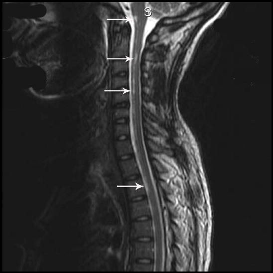
Figures

ill-defined central T2 hyperintensity is seen in axial view at mid T6-level
enlarged sagittal image of thoracic cord demonstrating central T2 hyperintensity from T5-T8 (arrows) and Schmorl’s node (arrowhead). Infarction down to T10 can be seen on other cuts.
References
-
- Han JJ, Massagli TL, Jaffe KM. Fibrocartilaginous embolism - an uncommon cause of spinal cord infarction: Case report and review of the literature. Arch Phys Med Rehabil. 2004;85:153–7. - PubMed
-
- Davis GA, Klug GL. Acute-onset nontraumatic paraplegia in childhood: Fibrocartilaginous embolism or acute myelitis? Child's Nervous System. 2000;16:551–4. - PubMed
-
- International Classification of Diseases (icd - 9 - cm) Denver, DE: American Medical Association; 1999.
Publication types
MeSH terms
Grants and funding
LinkOut - more resources
Full Text Sources
Medical
