. 2007 May 1;21(9):1025-30.
doi: 10.1101/gad.1540407. Epub 2007 Apr 16.

The tumor suppressor microRNA let-7 represses the HMGA2 oncogene

Affiliations

The tumor suppressor microRNA let-7 represses the HMGA2 oncogene

Yong Sun Lee et al. Genes Dev. .

Abstract

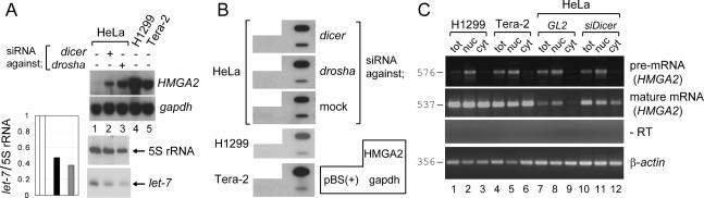
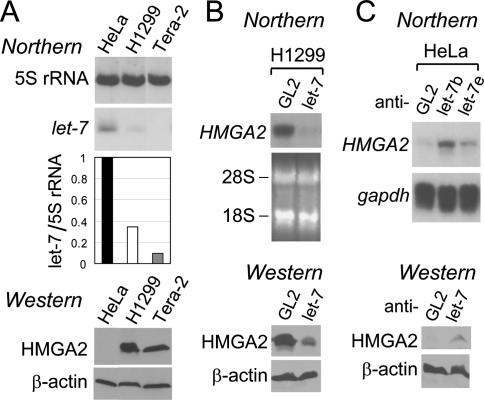
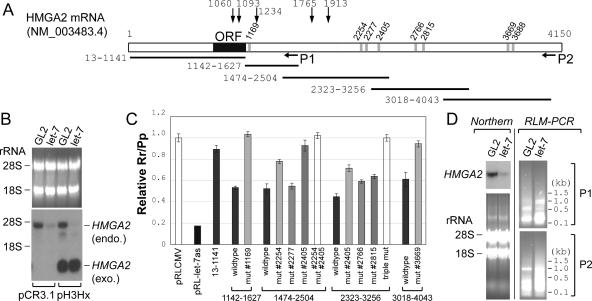
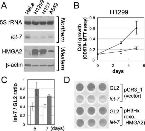
HMGA2, a high-mobility group protein, is oncogenic in a variety of tumors, including benign mesenchymal tumors and lung cancers. Knockdown of Dicer in HeLa cells revealed that the HMGA2 gene is transcriptionally active, but its mRNA is destabilized in the cytoplasm through the microRNA (miRNA) pathway. HMGA2 was derepressed upon inhibition of let-7 in cells with high levels of the miRNA. Ectopic expression of let-7 reduced HMGA2 and cell proliferation in a lung cancer cell. The effect of let-7 on HMGA2 was dependent on multiple target sites in the 3' untranslated region (UTR), and the growth-suppressive effect of let-7 on lung cancer cells was rescued by overexpression of the HMGA2 ORF without a 3'UTR. Our results provide a novel example of suppression of an oncogene by a tumor-suppressive miRNA and suggest that some tumors activate the oncogene through chromosomal translocations that eliminate the oncogene's 3'UTR with the let-7 target sites.


Figures

Figure 1.
Figure 1.
Destabilization of HMGA2 mRNA in the cytoplasm of HeLa cells through the miRNA pathway. (A, top two panels) Northern hybridization to detect HMGA2 and gapdh as a control. HeLa cells were transfected with GL2 (lane 1) or siRNA against Dicer (target sequence: AACUCUGCAAACCAGGUUGCU) or Drosha (target sequence: AACGAGUAGGCUUCGUGACUU) (lanes 2,3). (Lanes 4,5) The results from H1299 and Tera-2 cells are included for comparison. (Bottom two panels and the graph on the left) The levels of let-7 miRNA and 5S rRNA were measured by small RNA Northern. The blot was exposed to PhosphorImager Storm 860 (Molecular Dynamics), and each band was quantified with ImageQuant 5.2 software. The intensity of let-7 was normalized to that of 5S rRNA and plotted on the left of the blot (plain bar, lane 1; black bar, lane 2; gray bar, lane 3). (Lane 1) The value of the GL2 transfected sample was set as 1. (B) Nuclear run-on experiments were performed with the nuclei corresponding to the samples in A. The identity of each band is marked in a separate box on the right. pBS(+) indicates the pBluescript vector negative control. (C) To detect the pre-mRNA and mature mRNA of HMGA2, RT–PCR was performed with the RNAs from total (tot), nuclear (nuc), and cytoplasmic (cyt) fractions. Each amplified fragment with its expected size in base pairs is indicated. To ensure PCR products are clearly visible but are not saturated, we adjusted the amount of input cDNA and the number of cycles in each PCR amplification (35 cycles with fourfold more cDNA to detect the pre-mRNA of HMGA2, 30 cycles for mature mRNA of HMGA2, and 22 cycles for β-actin control). A representative result from the reaction without reverse transcriptase (−RT) is shown.
Figure 2.
Figure 2.
The effect of let-7 miRNA on HMGA2 expression. (A, top two panels and graph) let-7 and 5S rRNA from the indicated cell lines were measured as described in Figure 1A (black bar, HeLa; plain bar, H1299; gray bar, Tera-2). (Bottom two panels) Immunoblots for HMGA2 protein (using an antibody from Oxis International, Inc.) and β-actin as a loading control. (B, top two panels) After transfection of GL2 and let-7 duplex (a mixture of let-7b and let-7e) into H1299 lung cancer cells, HMGA2 mRNA was measured by Northern hybridization, with ethidium bromide staining of two ribosomal RNA bands shown as a loading control. (Bottom two panels) Immunoblots for HMGA2 and β-actin proteins. (C) HMGA2 mRNA (top two panels) and protein (bottom two panels) were detected after transfection of the indicated 2′-O-methyl antisense oligonucleotides into HeLa cells, with gapdh mRNA and β-actin protein as controls, respectively.
Figure 3.
Figure 3.
The destabilization of HMGA2 mRNA by let-7 is dependent on the predicted target sites in the 3′UTR. (A) A schematic of HMGA2 mRNA. All numbers are based on the nucleotide sequence of NM_ 003483.4. Vertical gray bars indicate the potential binding sites of let-7, predicted by miRanda, PICTAR, or TargetScan (for details, see Supplementary Table S2). The target sites are named according to the nucleotide coordinate of target sequence that pairs with the 5′ end of let-7. P1 and P2 indicate approximate locations of each set of primers for the RLM-PCR (shown in D) (for details, see also Supplementary Fig. S3 and Supplementary Table S2). Horizontal lines represent segments of HMGA2 used in the luciferase reporter assays in C. Vertical arrows with the nucleotide coordinates indicate locations of chromosomal breaks in human tumors (Schoenmakers et al. 1995; Geurts et al. 1997; Ashar et al. 2003; Quade et al. 2003; Nilsson et al. 2005; Odero et al. 2005). (B) Northern hybridization was performed with the H1299-derived cell line stably expressing HMGA2 from the plasmid pH3Hx or the control cell line containing the parental vector pCR3.1, as indicated at the bottom. Endogenous HMGA2 mRNA (endo.) and the HMGA2 mRNA lacking the 3′UTR (from pH3Hx; exo.) are indicated. The rest are as in the Northern blot in Figure 2B. (C) Repression of luciferase reporter by let-7 in H1299 cells. pRL-CMV derivatives contained the indicated segments of HMGA2 mRNA inserted downstream from the Renilla luciferase ORF (the numbers at the bottom match the horizontal lines in A). pRL-let-7as is a control plasmid containing a sequence perfectly complementary to let-7 downstream from the luciferase and is repressed by the let-7 duplex by siRNA mechanisms. The relative Rr/Pp value (Y-axis) indicates the extent to which let-7 represses a given reporter. The average and standard deviation of triplicate samples are indicated. (D) RLM-PCR was performed with RNA from H1299 cells at 24 h after transfection of GL2 or let-7 duplex. (Left two panels) Northern hybridization as described in Figure 2B. (Right two panels) Agarose gel electrophoresis of RLM-RACE products from primer set P1 and P2 (shown in A) (Supplementary Fig. S3; Supplementary Table S1), with molecular size markers (in kilobases) indicated.
Figure 4.
Figure 4.
Inhibition of proliferation of H1299 by let-7 is rescued by let-7-resistant HMGA2. (A) Decrease in let-7 and increase in HMGA2 in three lung cancer cell lines—H1299, H157, and A549. The rest are as described in Figure 2A. (B) The growth of H1299 under 2% serum was measured after transfection of GL2 (filled triangle) or let-7 duplex (dash). X-axis shows days after the first transfection; Y-axis shows OD at 570 nm in MTT assays. (C,D) Same experiment as in B was performed in H1299 stably expressing HMGA2 ORF (H1299 pH3Hx; gray bar) or containing the vector control (H1299 pCR3.1; plain bar). (Y-axis) The MTT value of cells transfected with let-7 relative to those transfected with the control duplex GL2. (D) A representative scan of the wells after the MTT assay at day 7.

Comment in

  • Oncogenic HMGA2: short or small?
    Young AR, Narita M. Young AR, et al. Genes Dev. 2007 May 1;21(9):1005-9. doi: 10.1101/gad.1554707. Genes Dev. 2007. PMID: 17473167 Review. No abstract available.

References

    1. Ashar H.R., Tkachenko A., Shah P., Chada K., Tkachenko A., Shah P., Chada K., Shah P., Chada K., Chada K. HMGA2 is expressed in an allele-specific manner in human lipomas. Cancer Genet. Cytogenet. 2003;143:160–168. - PubMed
    1. Ayoubi T.A., Jansen E., Meulemans S.M., de Van Ven W.J., Jansen E., Meulemans S.M., de Van Ven W.J., Meulemans S.M., de Van Ven W.J., de Van Ven W.J. Regulation of HMGIC expression: An architectural transcription factor involved in growth control and development. Oncogene. 1999;18:5076–5087. - PubMed
    1. Berlingieri M.T., Manfioletti G., Santoro M., Bandiera A., Visconti R., Giancotti V., Fusco A., Manfioletti G., Santoro M., Bandiera A., Visconti R., Giancotti V., Fusco A., Santoro M., Bandiera A., Visconti R., Giancotti V., Fusco A., Bandiera A., Visconti R., Giancotti V., Fusco A., Visconti R., Giancotti V., Fusco A., Giancotti V., Fusco A., Fusco A. Inhibition of HMGI-C protein synthesis suppresses retrovirally induced neoplastic transformation of rat thyroid cells. Mol. Cell. Biol. 1995;15:1545–1553. - PMC - PubMed
    1. Berner J.M., Meza-Zepeda L.A., Kools P.F., Forus A., Schoenmakers E.F., de Van Ven W.J., Fodstad O., Myklebost O., Meza-Zepeda L.A., Kools P.F., Forus A., Schoenmakers E.F., de Van Ven W.J., Fodstad O., Myklebost O., Kools P.F., Forus A., Schoenmakers E.F., de Van Ven W.J., Fodstad O., Myklebost O., Forus A., Schoenmakers E.F., de Van Ven W.J., Fodstad O., Myklebost O., Schoenmakers E.F., de Van Ven W.J., Fodstad O., Myklebost O., de Van Ven W.J., Fodstad O., Myklebost O., Fodstad O., Myklebost O., Myklebost O. HMGIC, the gene for an architectural transcription factor, is amplified and rearranged in a subset of human sarcomas. Oncogene. 1997;14:2935–2941. - PubMed
    1. Bol S., Wanschura S., Thode B., Deichert U., de Van Ven W.J., Bartnitzke S., Bullerdiek J., Wanschura S., Thode B., Deichert U., de Van Ven W.J., Bartnitzke S., Bullerdiek J., Thode B., Deichert U., de Van Ven W.J., Bartnitzke S., Bullerdiek J., Deichert U., de Van Ven W.J., Bartnitzke S., Bullerdiek J., de Van Ven W.J., Bartnitzke S., Bullerdiek J., Bartnitzke S., Bullerdiek J., Bullerdiek J. An endometrial polyp with a rearrangement of HMGI-C underlying a complex cytogenetic rearrangement involving chromosomes 2 and 12. Cancer Genet. Cytogenet. 1996;90:88–90. - PubMed

MeSH terms

LinkOut - more resources