. 2007 Jul;75(7):3256-63.
doi: 10.1128/IAI.01984-06. Epub 2007 Apr 16.

A functional NADPH oxidase prevents caspase involvement in the clearance of phagocytic neutrophils

Affiliations

A functional NADPH oxidase prevents caspase involvement in the clearance of phagocytic neutrophils

Rachel P Wilkie et al. Infect Immun. 2007 Jul.

Abstract

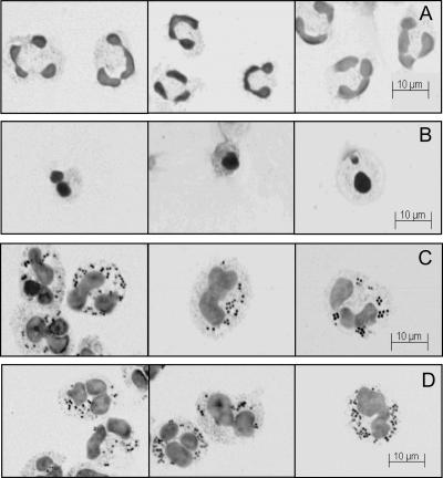
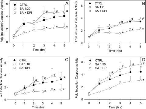
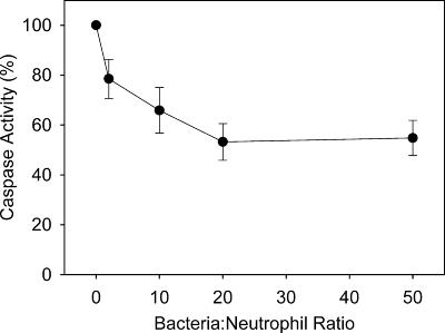
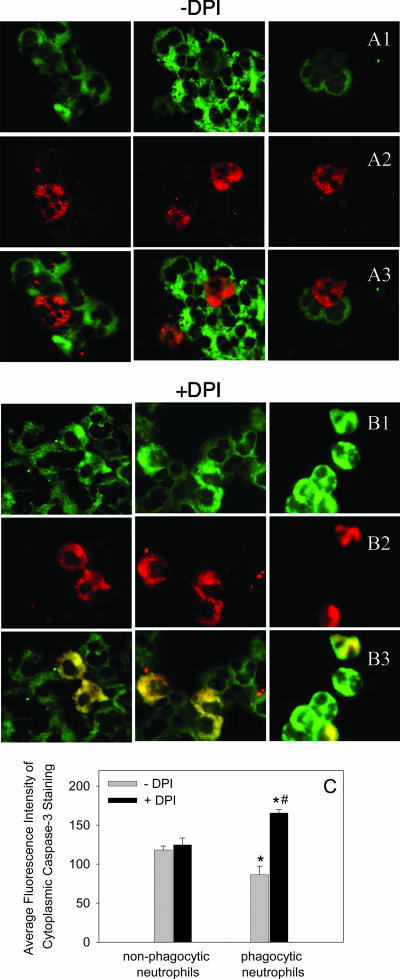
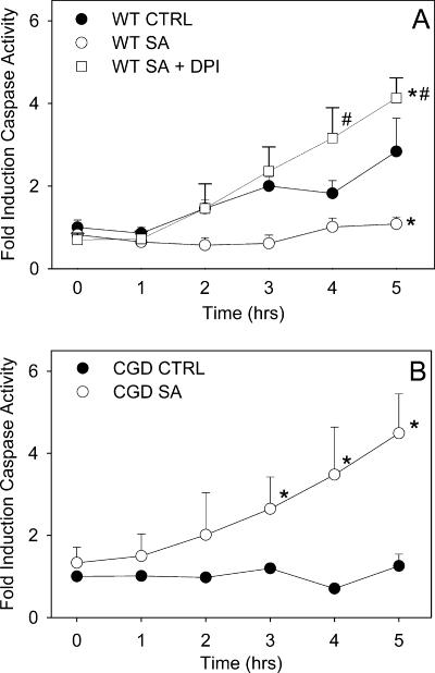
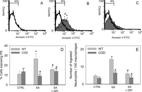
Neutrophils play a prominent role in host defense. Phagocytosis of bacteria leads to the formation of an active NADPH oxidase complex that generates reactive oxygen species for bactericidal purposes. A critical step in the resolution of inflammation is the uptake of neutrophils by macrophages; however, there are conflicting reports on the mechanisms leading to the apoptosis of phagocytic neutrophils. The aim of this study was to clarify the role of effector caspases in these processes. Caspase activity was measured by DEVDase activity assays or immunofluorescence detection of active caspase-3. With normal human and wild-type murine neutrophils there was no caspase activation following phagocytosis of Staphylococcus aureus. However, caspase activity was observed in phagocytic neutrophils with a defective NADPH oxidase, including neutrophils isolated from X-linked gp91(phox) knockout chronic granulomatous disease mice. These results indicate that a functional NADPH oxidase and the generation of oxidants in the neutrophil phagosome prevent the activation of the cytoplasmic caspase cascade.


Figures

FIG. 1.
FIG. 1.
Phagocytosis of S. aureus by human neutrophils and subsequent changes in morphology. Photographs from a representative experiment for cytospins of human unstimulated neutrophils at 3 h (A), of untreated neutrophils after 48 h of incubation (B), of neutrophils with S. aureus at 3 h (C), and of neutrophils with S. aureus at 5 h (D).
FIG. 2.
FIG. 2.
Caspase-3 activation is blocked by NADPH oxidase activity in phagocytic human neutrophils. Caspase activity was assessed hourly in neutrophils incubated alone (CTRL), in neutrophils incubated with S. aureus (SA) at ratios of 1:20 (A), 1:2 (B), 1:10 (C), and 1:50 (D), and in neutrophils treated with DPI prior to coincubation with S. aureus (SA + DPI). The active caspase-3 activity was measured by monitoring the arbitrary units of fluorescence liberated following cleavage of the fluorogenic peptide substrate DEVD-AMC. The means and standard errors of the means of four to nine experiments are shown. An asterisk indicates that the P value is <0.05 for a comparison with the control, and a number sign indicates that the P value is <0.05 for a comparison with phagocytic neutrophils with a functional NADPH oxidase.
FIG. 3.
FIG. 3.
Dose-dependent inhibition of caspase activity. Caspase activity in neutrophils incubated with S. aureus at ratios of 1:2, 1:10, 1:20, and 1:50 after 1 h of incubation is expressed as a percentage of the activity expressed at 1 h in unstimulated neutrophils.
FIG. 4.
FIG. 4.
Immunofluorescence staining for active caspase-3. Neutrophils were incubated either alone or with PI-labeled S. aureus (1:20) for 3 h, and then both phagocytic and nonphagocytic neutrophils were mixed and cytospun onto the same microscope slide. Cells were treated with a cleaved caspase-3 antibody, followed by a secondary antibody conjugated to FITC, and were visualized with a fluorescent microscope. Images of the green fluorescence (A1 and B1) showing positive caspase-3 staining localized in the cytosol and the red fluorescence (A2 and B2) identifying phagocytic neutrophils were captured from the same field, and the pairs of images were superimposed (A3 and B3). Untreated phagocytic neutrophils (A2) surrounded by nonphagocytic neutrophils (A3) show negligible caspase-3 activity compared to the activity of the neighboring nonphagocytic cells (A1). When phagocytic neutrophils were pretreated with DPI (B1 to B3) to inhibit the NADPH oxidase, the phagocytic cells adjacent to nonphagocytic neutrophils (B3) displayed enhanced caspase activation (B1). The photographs are from representative experiments. (C) Quantification of caspase-3 fluorescence within untreated and phagocytic neutrophils with or without DPI treatment, analyzed using the Discovery-1 high-throughput and high-content screening machine. Fluorescence intensity was assessed in 300 cells from 27 photographs taken in three different experiments. An asterisk indicates that the P value is <0.05 for a comparison with the control, and a number sign indicates that the P value is <0.05 for a comparison with phagocytic neutrophils with a functional NADPH oxidase.
FIG. 5.
FIG. 5.
Caspase-3 activation in phagocytic murine neutrophils. Neutrophils were incubated either alone (CTRL) or with S. aureus (SA) (1:20) or were treated with DPI prior to coincubation with S. aureus (SA + DPI). Caspase-3 activity was assessed hourly by monitoring the increase in fluorescence with excitation at 390 nm and emission at 460 nm following cleavage of the fluorogenic peptide substrate DEVD-AMC. The means and standard errors of the means of 3 to 10 experiments are shown. An asterisk indicates that the P value is <0.05 for a comparison with the control, and a number sign indicates that the P value is <0.05 for a comparison with phagocytic neutrophils with a functional NADPH oxidase. WT, wild type.
FIG. 6.
FIG. 6.
Phagocytosis triggers PS exposure and uptake by macrophages in murine neutrophils and requires a functional NADPH oxidase. After 4 h of incubation with S. aureus (1:20) cells were stained with annexin V-FITC and PI (A to C). The flow cytometry histograms show the results of a single representative experiment with mouse wild-type neutrophils. (A) Unstimulated neutrophils; (B) neutrophils with S. aureus (shaded area); (C) DPI-treated neutrophils with S. aureus (shaded area). The results of treatment of neutrophils with S. aureus in the presence of the caspase inhibitor z-VAD-fmk are indicated by the dashed line in panel B. (D) Percentage of murine wild-type (WT) and CGD neutrophils exposing PS following incubation alone (CTRL), following incubation with S. aureus (SA), or following pretreatment with DPI (SA + DPI). (E) For macrophage uptake studies, murine neutrophils were incubated with S. aureus, harvested, and layered onto monocyte-derived macrophages, the medium was removed, and the wells were fixed and stained for myeloperoxidase with o-dianisidine, enabling visualization of neutrophils. The number of neutrophils phagocytosed per 100 macrophages was determined. The means and standard errors of the means of three to eight experiments are shown. An asterisk indicates that the P value is <0.05 for a comparison with the control, and a number sign indicates that the P value is <0.05 for a comparison with phagocytic neutrophils with a functional NADPH oxidase.

References

    1. Baran, J., K. Guzik, W. Hryniewicz, M. Ernst, H. D. Flad, and J. Pryjma. 1996. Apoptosis of monocytes and prolonged survival of granulocytes as a result of phagocytosis of bacteria. Infect. Immun. 64:4242-4248. - PMC - PubMed
    1. Borjesson, D. L., S. D. Kobayashi, A. R. Whitney, J. M. Voyich, C. M. Argue, and F. R. Deleo. 2005. Insights into pathogen immune evasion mechanisms: Anaplasma phagocytophilum fails to induce an apoptosis differentiation program in human neutrophils. J. Immunol. 174:6364-6372. - PubMed
    1. Brach, M. A., S. deVos, H. J. Gruss, and F. Herrmann. 1992. Prolongation of survival of human polymorphonuclear neutrophils by granulocyte-macrophage colony-stimulating factor is caused by inhibition of programmed cell death. Blood 80:2920-2924. - PubMed
    1. Cheah, F. C., M. B. Hampton, B. A. Darlow, C. C. Winterbourn, and M. C. Vissers. 2005. Detection of apoptosis by caspase-3 activation in tracheal aspirate neutrophils from premature infants: relationship with NF-kappaB activation. J. Leukoc. Biol. 77:432-437. - PubMed
    1. Colotta, F., F. Re, N. Polentarutti, S. Sozzani, and A. Mantovani. 1992. Modulation of granulocyte survival and programmed cell death by cytokines and bacterial products. Blood 80:2012-2020. - PubMed

Publication types

MeSH terms

LinkOut - more resources