Comparative Study
. 2007 May 14;204(5):1049-56.
doi: 10.1084/jem.20062566. Epub 2007 Apr 16.

Fetal gene defects precipitate platelet-mediated pregnancy failure in factor V Leiden mothers

Affiliations
Comparative Study

Fetal gene defects precipitate platelet-mediated pregnancy failure in factor V Leiden mothers

Rashmi Sood et al. J Exp Med. .

Abstract

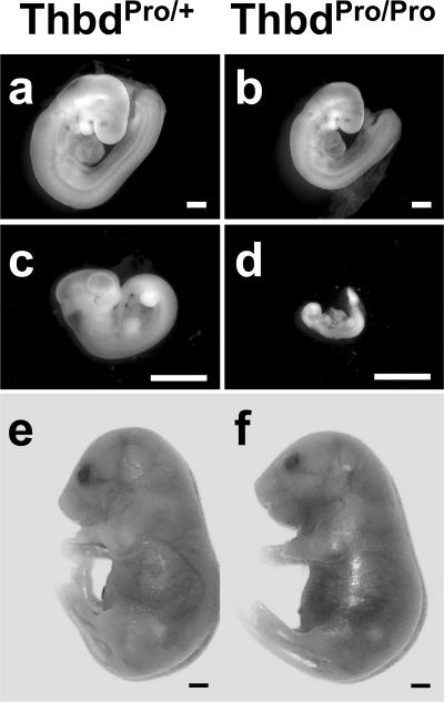
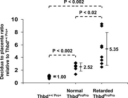
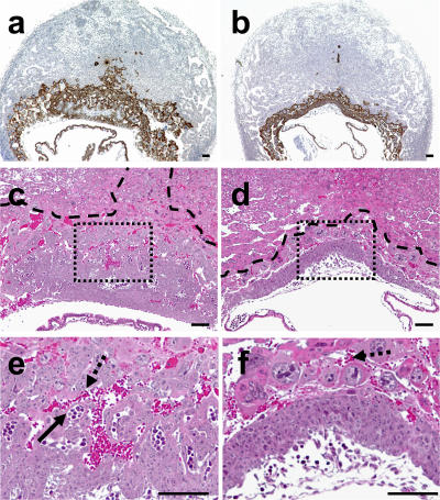
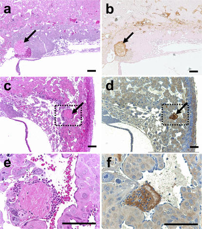
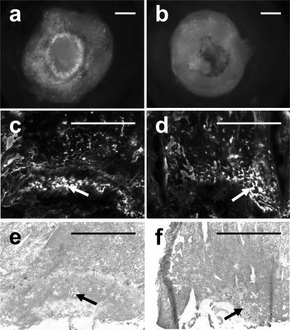
We describe a mouse model of fetal loss in factor V Leiden (FvL) mothers in which fetal loss is triggered when the maternal prothrombotic state coincides with fetal gene defects that reduce activation of the protein C anticoagulant pathway within the placenta. Fetal loss is caused by disruption of placental morphogenesis at the stage of labyrinth layer formation and occurs in the absence of overt placental thrombosis, infarction, or perfusion defects. Platelet depletion or elimination of protease-activated receptor 4 (Par4) from the mother allows normal placentation and prevents fetal loss. These findings establish a cause-effect relationship for the observed epidemiologic association between maternal FvL status and fetal loss and identify fetal gene defects as risk modifiers of pregnancy failure in prothrombotic mothers. Pregnancy failure is mediated by Par4-dependent activation of maternal platelets at the fetomaternal interface and likely involves a pathogenic pathway independent of occlusive thrombosis. Our results further demonstrate that the interaction of two given thrombosis risk factors produces markedly disparate consequences on disease manifestation (i.e., thrombosis or pregnancy loss), depending on the vascular bed in which this interaction occurs.


Figures

Figure 1.
Figure 1.
Phenotype of ThbdProPro embryos in FVQQThbdPro+ mothers. (a, c, and e) ThbdPro+ littermates at E9.5, E12.5, and E18.5, respectively, of ThbdProPro embryos (b, d, and f). At E9.5, some ThbdProPro embryos are smaller (b). By E11.5, most ThbdProPro embryos are in an advanced stage of decay (d). Platelet depletion of the mother with anti-GP1bα antibody treatment (e and f) results in normal-appearing ThbdProPro embryos. Pictures were taken at different magnifications. Bars, 1 mm.
Figure 2.
Figure 2.
Placental size of ThbdProPro embryos relative to controls. Each symbol represents a measurement from a single fetoplacental unit. All litters were analyzed at E9.5. Mean ratios and SDs are shown.
Figure 3.
Figure 3.
Placental phenotype of ThbdProPro embryos and littermate controls in FvQQThbdPro+ mothers. Histological sections through placentas of ThbdPro+ (a, c, and e) and a phenotypically normal ThbdProPro (b, d, and f) embryo at E9.5. a and b show cytokeratin-expressing trophoblast cells (brown staining). The placenta of ThbdProPro embryos (stained area) is smaller than that of ThbdPro+ embryos. c and d are higher magnification images of hematoxylin and eosin–stained sections adjacent to a and b, respectively. e and f correspond to boxes in c and d, respectively. Dashed lines in c and d mark the border of the placenta and decidua, as determined by cytokeratin staining. ThbdPro+ placentas (c and e) have formed a labyrinth characterized by maternal (bright red enucleated maternal red blood cells; dashed arrows) and fetal (purple hematoxylin-stained nucleated fetal red blood cells; continuous arrow) blood spaces. The labyrinth is distinctly absent in the placentas of littermate ThbdProPro embryos (d and f). Bars, 0.1 mm.
Figure 4.
Figure 4.
Placental thrombosis in FvL mice. The number of placental thrombi was similar in placentas of wild-type and ThbdProPro embryos (see Results). Thrombi were identified on serial sections by hematoxylin and eosin staining (a, c, and e), and immunostaining with antifibrinogen antibodies (b) and anti–p-selectin antibodies (d and f). e and f are enlarged views of the boxed areas in c and d. Arrows indicate clots. Bars, 0.1 mm.
Figure 5.
Figure 5.
Assessment of decidual and placental vascular patency by FITC-dextran infusion. Whole-mount (a and b) and sections (c–f) of decidual-placental units corresponding to a wild-type embryo in a wild-type mother (a, c, and e) and a FvQ+ThbdProPro embryo in a FvQQThbdPro+ mother (b, d, and f) are shown. From the fetal aspect of the placenta, a characteristic ring of maternal placental vasculature, marked by FITC-dextran, is observed in wild-type pregnancies but not in placenta of a FvQ+ThbdProPro embryo in a FvQQThbdPro+ mother (a and b). Decidual and placental regions in both types of pregnancies are perfused with FITC-dextran (c and d). Hematoxylin and eosin–stained sections adjacent to c and d are shown in e and f. Arrows show maternal blood spaces in the placenta. Bars, 1 mm.

Similar articles

Cited by

References

    1. Dudding, T.E., and J. Attia. 2004. The association between adverse pregnancy outcomes and maternal factor V Leiden genotype: a meta-analysis. Thromb. Haemost. 91:700–711. - PubMed
    1. Rey, E., S.R. Kahn, M. David, and I. Shrier. 2003. Thrombophilic disorders and fetal loss: a meta-analysis. Lancet. 361:901–908. - PubMed
    1. Kovalevsky, G., C.R. Gracia, J.A. Berlin, M.D. Sammel, and K.T. Barnhart. 2004. Evaluation of the association between hereditary thrombophilias and recurrent pregnancy loss: a meta-analysis. Arch. Intern. Med. 164:558–563. - PubMed
    1. Mousa, H.A., and Z. Alfirevic. 2000. Do placental lesions reflect thrombophilia state in women with adverse pregnancy outcome? Hum. Reprod. 15:1830–1833. - PubMed
    1. Sikkema, J.M., A. Franx, H.W. Bruinse, N.G. van der Wijk, H.W. de Valk, and P.G. Nikkels. 2002. Placental pathology in early onset pre-eclampsia and intra-uterine growth restriction in women with and without thrombophilia. Placenta. 23:337–342. - PubMed

Publication types

MeSH terms