. 2007 Apr 23;177(2):289-303.
doi: 10.1083/jcb.200609072. Epub 2007 Apr 16.

A signal from inside the peroxisome initiates its division by promoting the remodeling of the peroxisomal membrane

Affiliations

A signal from inside the peroxisome initiates its division by promoting the remodeling of the peroxisomal membrane

Tong Guo et al. J Cell Biol. .

Abstract

We define the dynamics of spatial and temporal reorganization of the team of proteins and lipids serving peroxisome division. The peroxisome becomes competent for division only after it acquires the complete set of matrix proteins involved in lipid metabolism. Overloading the peroxisome with matrix proteins promotes the relocation of acyl-CoA oxidase (Aox), an enzyme of fatty acid beta-oxidation, from the matrix to the membrane. The binding of Aox to Pex16p, a membrane-associated peroxin required for peroxisome biogenesis, initiates the biosynthesis of phosphatidic acid and diacylglycerol (DAG) in the membrane. The formation of these two lipids and the subsequent transbilayer movement of DAG initiate the assembly of a complex between the peroxins Pex10p and Pex19p, the dynamin-like GTPase Vps1p, and several actin cytoskeletal proteins on the peroxisomal surface. This protein team promotes membrane fission, thereby executing the terminal step of peroxisome division.


Figures
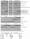
Figure 1.
Figure 1.
Pex16p regulates lipid metabolism in the peroxisomal membrane. (A) Spectra of membrane lipids in different peroxisomal subforms purified from wild-type, pex16Δ, and PEX16-TH cells. Peroxisomes were osmotically lysed and subjected to centrifugation. Lipids were extracted from equal quantities of the pelleted membrane proteins and analyzed by TLC. (B and C) Dynamics of radiolabeled lipids in the membrane of P1 liposomes. Liposomes were reconstituted from PMPs immunodepleted (−Pex16p) or not immunodepleted (+Pex16p) of Pex16p and from nonradiolabeled lipids, all of which were extracted from the membrane of immature peroxisomal vesicles P1. [14C]LPA (B) or [14C]PA (C) were the only radiolabeled lipids incorporated into liposomes during their reconstitution. The [14C]LPA-loaded liposomes (B) were also supplemented with unlabeled oleoyl-CoA, a cosubstrate of LPAAT. Samples were taken at the indicated times after transfer of reconstituted liposomes from ice to 26oC. Lipids were extracted from the membrane and analyzed by TLC. (D) Pex16p inhibits LPAAT, the first enzyme in a two-step biosynthetic pathway leading to the formation of DAG in the peroxisomal membrane during conversion of P5 to P6.
Figure 2.
Figure 2.
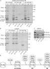
In all six peroxisomal subforms, both LPAAT and PAP are integral membrane proteins that do not face the cytosol and are crucial for the biosynthesis of PA and DAG in the peroxisomal membrane. (A) Purification of LPAAT and PAP from the membrane of mature peroxisomes P6. Peroxisomes of wild-type strain were osmotically lysed and subjected to centrifugation. Pelleted membrane proteins were extracted with Na+ cholate and subjected to a series of the chromatographic purification steps, as indicated. Proteins recovered in the Na+ cholate-extracted membrane of P6 and in chromatographic fractions were resolved by SDS-PAGE, followed by silver staining. For monitoring enzymatic activities of LPAAT and PAP, recovered membrane proteins were incorporated into the membrane of peroxisomal liposomes that were reconstituted from the PMPs and membrane lipids of P6. [14C]-labeled lipid substrates of LPAAT and PAP were incorporated into liposomes as described in the legend to Fig. 1. The spectra of proteins and enzymatic activities of LPAAT and PAP recovered in the Na+ cholate–extracted membrane of P6 and in the peak chromatographic fractions are shown. Arrowheads mark LPAAT and PAP, which were purified to apparent homogeneity and identified by mass spectrometry as the Slc1p and Dpp1p proteins, respectively. (B) Peroxisomal subforms P1–P6 were purified from wild-type (wt) cells. Equal quantities (10 μg) of protein from these peroxisomes, as well as equal quantities (100 μg) of protein from lysates of whole cells of wild-type, slc1Δ, and dpp1Δ strains, were analyzed by immunoblotting with antibodies to Slc1p (LPAAT) and Dpp1p (PAP). (C) Equal aliquots (10 μg of total protein) of mature peroxisomes purified from wild-type cells were osmotically lysed or exposed to 1 M NaCl, 0.1 M Na2CO3, pH 11.0, or 0.5% (vol/vol) Triton X-100. After incubation on ice for 30 min, samples were separated into supernatant (S) and pellet (P) fractions by centrifugation and then immunoblotted with the indicated antibodies. (D) Equal aliquots (10 μg of total protein) of mature peroxisomes purified from wild-type cells were treated with the indicated amounts of trypsin in the absence (−) or presence (+) of 0.5% (vol/vol) Triton X-100 for 30 min on ice. Samples were subjected to SDS-PAGE and immunoblotting with the indicated antibodies. (E) Spectra of membrane lipids in different peroxisomal subforms purified from wild-type, slc1Δ, and dpp1Δ cells. Peroxisomes were osmotically lysed and subjected to centrifugation. Lipids were extracted from equal quantities of the pelleted membrane proteins and analyzed by TLC.
Figure 3.
Figure 3.
Pex16p binds to LPA only in the membranes of division-incompetent peroxisomal subforms. Different peroxisomal subforms purified from wild-type cells (A) and highly purified mature peroxisomes P6 of wild-type and mutant strains (B) were osmotically lysed and subjected to centrifugation. The pellet of membranes after such centrifugation was solubilized with a detergent, n-OG. Equal quantities of detergent-soluble membrane proteins were analyzed by protein-lipid overlay assay using commercial PIP Strips. Pex16p was detected by immunoblotting with anti-Pex16p antibodies.
Figure 4.
Figure 4.
Mutations that abolish the binding of Aox to Pex16p, thereby impairing peroxisome division, prevent the biosynthesis of PA and DAG in the peroxisomal membrane. Highly purified peroxisomal subforms were osmotically lysed and subjected to centrifugation. Equal quantities of the pelleted membrane proteins recovered from different peroxisomal subforms were subjected to lipid extraction, which was followed by TLC and visualization of lipids.
Figure 5.
Figure 5.
PC in the peroxisomal membrane is a positive regulator of both LPAAT and PAP. (A and B) The initial rates of the LPAAT (A) and PAP (B) reactions and the levels of PC recovered in the membranes of liposomes reconstituted from the Pex16p-immunodepleted PMPs and membrane lipids of different peroxisomal subforms. Peroxisomal liposomes that lack Pex16p were reconstituted as described in the legend to Fig. 1. [14C]-labeled lipid substrates were incorporated into liposomes during their reconstitution. (C–F) The initial rates of the LPAAT (C and E) and PAP (D and F) reactions and the levels of PC recovered in the membranes of four different types of liposomes reconstituted from the Pex16p-immunodepleted PMPs and membrane lipids of P1 (C– F), P2 (E and F), or P3 (E and F) peroxisomes. These four different types of P1-, P2-, or P3-based liposomes varied only in the quantities of PC used for their reconstitution and recovered in their membranes after the reconstitution. For comparison, the initial rates of the LPAAT (E) and PAP (F) reactions and the levels of PC recovered in the membranes of liposomes reconstituted from the Pex16p-immunodepleted PMPs and membrane lipids of P4, P5, and P6 peroxisomes are shown. To calculate the initial rates of the LPAAT and PAP reactions, the [14C]-labeled LPA, PA, and DAG were separated by TLC and quantified by autoradiography. To visualize nonradiolabeled PC, lipids were separated by TLC and detected using phosphomolybdic acid.
Figure 6.
Figure 6.
A multiprotein complex that comprises a dynamin-like GTPase, three components of actin cytoskeleton, and two peroxins is assembled on the surface of the division-competent mature peroxisome. (A) Highly purified mature peroxisomes of wild-type (wt) and mutant strains were osmotically lysed and subjected to centrifugation to yield supernatant (matrix proteins) and pellet (membrane proteins) fractions. Recovered membrane proteins were treated with the thiol-cleavable cross-linker DSP. These DSP-treated membrane proteins were immunoprecipitated with anti-Vps1p, anti-Pex10p, and anti-Pex19p antibodies under denaturing, nonreducing conditions. The cross-linker was then cleaved with DTT, and the immunoprecipitated proteins were resolved by SDS-PAGE under reducing conditions, followed by silver staining. (B) Wild-type and mutant cells were subjected to subcellular fractionation to yield the 200S (cytosolic) fraction. Cytosolic proteins were treated with DSP. These DSP-treated cytosolic proteins were subjected to immunoprecipitation with anti-Vps1p and anti-Pex19p antibodies under denaturing, nonreducing conditions. The cross-linker was then cleaved with DTT, and the immunoprecipitated proteins were resolved by SDS-PAGE under reducing conditions, followed by silver staining. Arrows in A and B indicate the positions of Sla1p, Vps1p, Abp1p, Pex19p, Act1p, and Pex10p, which were identified by mass spectrometry. (C) Equal quantities (20 μg) of protein from mature peroxisomes of wild-type and mutant strains were analyzed by immunoblotting with the indicated antibodies. (D) A model for the multistep assembly of the Act1p–Abp1p–Sla1p–Vps1p–Pex19p–Pex10p complex on the surface of mature peroxisomes.
Figure 7.
Figure 7.
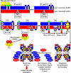
The Pex16p- and Aox-dependent intraperoxisomal signaling cascade drives the division of mature peroxisomes P6 by promoting the stepwise remodeling of lipid and protein composition of the peroxisomal membrane. The peroxisome becomes competent for division only after it acquires the complete set of matrix proteins involved in lipid metabolism. Overloading the peroxisome with matrix proteins promotes the relocation of Aox, an enzyme of fatty acid β-oxidation, from the matrix to the membrane. The binding of Aox to Pex16p, a membrane-associated peroxin required for peroxisome biogenesis, activates the biosynthesis and transbilayer movement of a distinct set of membrane lipids. The resulting remodeling of the lipid repertoire of the membrane bilayer initiates the stepwise assembly of a multicomponent protein complex on the surface of the mature peroxisome. This newly assembled protein complex carries out membrane fission, thereby executing the terminal step of peroxisome division.

Similar articles

Cited by

References

    1. Athenstaedt, K., and G. Daum. 1999. Phosphatidic acid, a key intermediate in lipid metabolism. Eur. J. Biochem. 266:1–16. - PubMed
    1. Bankaitis, V.A. 2002. Slick recruitment to the Golgi. Science. 295:290–291. - PubMed
    1. Behnia, R., and S. Munro. 2005. Organelle identity and the signposts for membrane traffic. Nature. 438:597–604. - PubMed
    1. Boukh-Viner, T., T. Guo, A. Alexandrian, A. Cerracchio, C. Gregg, S. Haile, R. Kyskan, S. Milijevic, D. Oren, J. Solomon, et al. 2005. Dynamic ergosterol- and ceramide-rich domains in the peroxisomal membrane serve as an organizing platform for peroxisome fusion. J. Cell Biol. 168:761–773. - PMC - PubMed
    1. Carman, G.M., and G.S. Han. 2006. Roles of phosphatidate phosphatase enzymes in lipid metabolism. Trends Biochem. Sci. 31:694–699. - PMC - PubMed

Publication types

MeSH terms