. 2007 Jun;27(12):4416-30.
doi: 10.1128/MCB.02314-06. Epub 2007 Apr 16.

cPGES/p23 is required for glucocorticoid receptor function and embryonic growth but not prostaglandin E2 synthesis

Affiliations

cPGES/p23 is required for glucocorticoid receptor function and embryonic growth but not prostaglandin E2 synthesis

Alysia Kern Lovgren et al. Mol Cell Biol. 2007 Jun.

Abstract

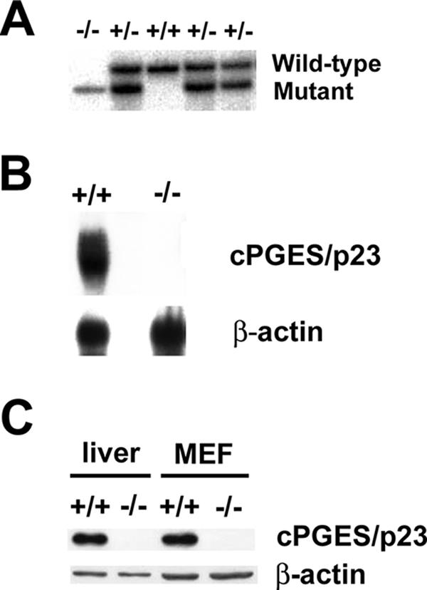
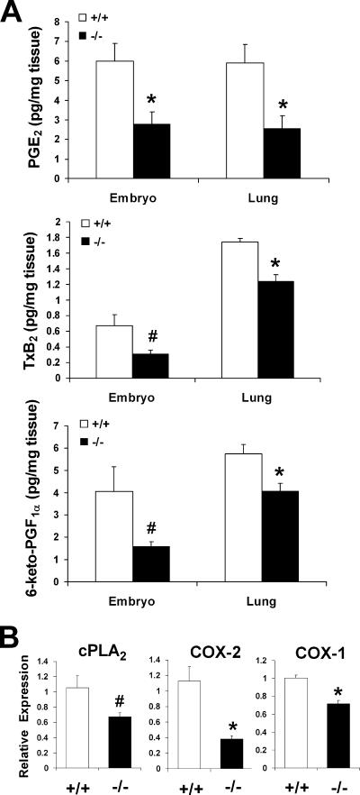
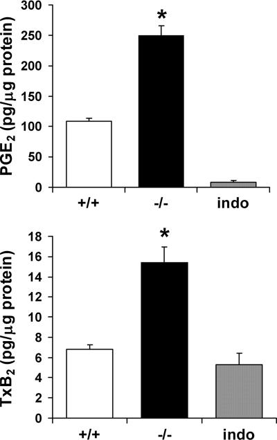
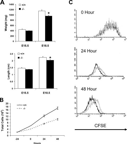
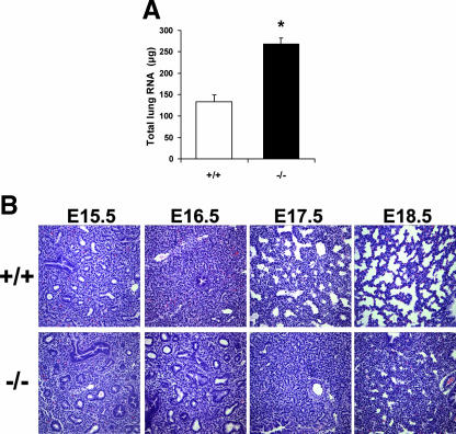
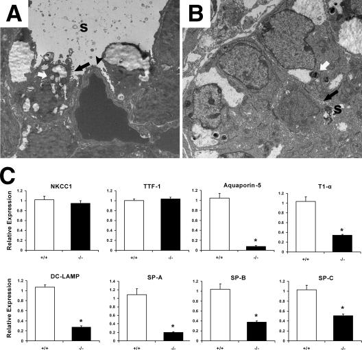
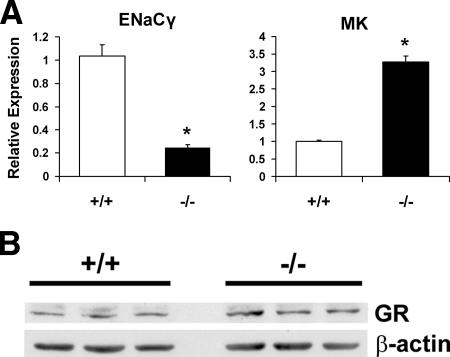
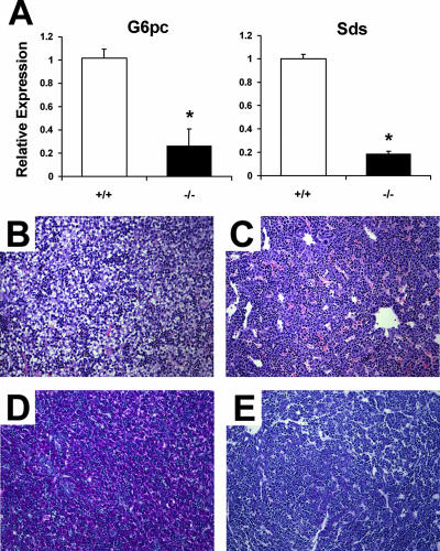
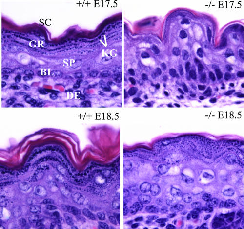
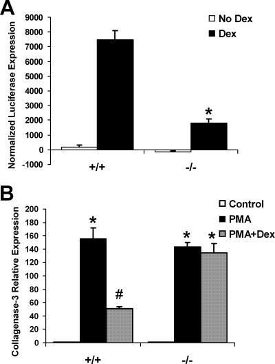
A number of studies have identified cytosolic prostaglandin E(2) synthase (cPGES)/p23 as a cytoplasmic protein capable of metabolism of prostaglandin E(2) (PGE(2)) from the cyclooxygenase metabolite prostaglandin endoperoxide (PGH(2)). However, this protein has also been implicated in a number of other pathways, including stabilization of the glucocorticoid receptor (GR) complex. To define the importance of the functions assigned to this protein, mice lacking detectible cPGES/p23 expression were generated. cPGES/p23(-/-) pups die during the perinatal period and display retarded lung development reminiscent of the phenotype of GR-deficient neonates. Furthermore, GR-sensitive gluconeogenic enzymes are not induced in the prenatal period. However, unlike GR-deficient embryos, cPGES/p23(-/-) embryos are small and a proliferation defect is observed in cPGES/p23(-/-) fibroblasts. Analysis of arachidonic acid metabolites in embryonic tissues and primary fibroblasts failed to support a function for this protein in PGE(2) biosynthesis. Thus, while the growth retardation of the cPGES/p23(-/-) pups and decreased proliferation of primary fibroblasts identify functions for this protein in addition to GR stabilization, it is unlikely that these functions include metabolism of PGH(2) to PGE(2).


Figures

FIG. 1.
FIG. 1.
Generation of cPGES/p23−/− mice. (A) Southern blot analyses of DNA from E18.5 embryos generated by the intercross of mice heterozygous for the cPGES/p23 mutant allele. (B) Northern blot analyses of cPGES/p23 mRNA expression in whole E18.5 embryos. Analysis with a β-actin-specific probe verified equal RNA sample loading. (C) Western blot analysis of cPGES/p23 protein expression in E18.5 liver and MEFs. β-Actin expression is shown as a loading control.
FIG. 2.
FIG. 2.
cPGES/p23-deficient embryos have a significant decrease in prostanoid levels and a corresponding decrease in cPLA2 and COX expression. (A) PGE2, TxB2, and 6-keto-PGF (a stable metabolite of prostacyclin [PGI2]) levels in whole E18.5 embryos and lungs dissected from E18.5 embryos were measured by enzyme immunoassay. n = 4. *, P < 0.05, and #, P ≤ 0.09 by Student's t test or P ≤ 0.05 by Mann-Whitney test. (B) Determination of cPLA2, COX-2, and COX-1 expression in total RNA isolated from E18.5 lungs by RT-PCR. Expression levels were normalized to β-actin, an endogenous control, and the results were expressed as change (fold) relative to wild-type expression levels. cPGES/p23−/− lungs, n = 7; wild-type lungs, n = 9. *, P < 0.05; #, P = 0.06.
FIG. 3.
FIG. 3.
cPGES/p23-deficient primary fibroblasts have a significant increase in PGE2 and TxB2 levels. MEFs were isolated from cPGES/p23−/− and wild-type embryos. Cells were seeded in 24-well plates and grown to 80% confluence. After 1 h of incubation in serum-free medium, the supernatant was removed and PGE2 and TxB2 levels in the supernatant were determined by enzyme immunoassay. The cells in each well were harvested for total protein determination by the BCA method. Results are expressed as picograms of prostanoid per microgram of total cell protein per well. Parallel cultures treated with indomethacin, which inhibits the enzymatic activities of both COX-1 and COX-2, served as a negative control in this assay. n = 6. *, P < 0.01.
FIG. 4.
FIG. 4.
Loss of cPGES/p23 results in growth defects in embryos and proliferation defects in the primary embryonic fibroblasts. (A) E15.5 and E18.5 embryos were removed by cesarean section, and weight and crown-to-rump length were determined. E15.5, cPGES/p23−/−, n = 6, and wild type, n = 4; E18.5, cPGES/p23−/−, n = 6, and wild type, n = 7. *, P < 0.05. (B) Growth curve for cPGES/p23−/− and wild-type primary embryonic fibroblasts. Equal numbers of cells of each genotype were plated in a six-well plate (three wells per time point for each genotype). At 24-h intervals, cells were harvested and counted. The supernatant was collected from each well, and the numbers of dead cells present in the supernatant did not differ between samples. In contrast, a marked reduction in the number of cells present in the wells seeded with cPGES/p23−/− cells was observed. These data are representative of two independent experiments, each performed with two different −/− and +/+ MEF cultures. (C) Proliferation assay comparing cPGES/p23−/− and wild-type primary fibroblasts. Cells were stained with CFSE and analyzed by flow cytometry 24 and 48 h after staining. The CFSE intensity at the time of labeling is shown at 0 h. A shift left at 24 and 48 h represents a decrease in CFSE intensity which corresponds to the rate of proliferation in the population. Shown are the representative results from three independent experiments. Black, wild type; gray, cPGES/p23−/−.
FIG. 5.
FIG. 5.
Histological analysis of lung tissue from E15.5 to E18.5 wild-type and cPGES/p23−/− embryos by light microscopy. (A) Total RNA was extracted from E18.5 embryo lungs. Quantitation of RNA revealed a significant increase in the cPGES/p23−/− lungs, suggesting hypercellularity. (B) Lungs were removed from embryos at E15.5, E16.5, E17.5, and E18.5 and fixed overnight in 10% formalin. Lung sections were stained with hematoxylin-eosin. The cPGES/p23−/− lung development appears arrested at the canalicular stage (E16.0 to 17.5). Notable is the failure of the development of terminal sacs. Original magnification, ×10.
FIG. 6.
FIG. 6.
Abnormal ultrastructure of the distal airway in cPGES/p23−/− embryos. Transmission electron microscopy was used to analyze ultrastructural morphology in E18.5 lung tissue from wild-type (A) and cPGES/p23−/− (B) embryos. The electron micrographs demonstrate squamous type I AECs (black arrowhead) and cuboidal type II AECs containing lamellar bodies (white arrows) and apical microvilli (black arrows) in the wild-type lung. In contrast, while type II AECs were detected in the cPGES/p23−/− lungs, type I AECs could not be identified. s, surfactant. (C) RT-PCR analysis was used to determine expression of a bronchial epithelial cell marker (NKCC1), a Clara cell-specific marker (TTF-1), type I AEC markers (aquaporin-5 and T1α), and type II AEC markers (DC-LAMP, SP-A, SP-B, and SP-C). Expression levels were normalized to β-actin, an endogenous control; the results are expressed as change (fold) relative to wild-type expression levels. cPGES/p23−/− lungs, n = 7; wild-type lungs, n = 9. *, P < 0.05.
FIG. 7.
FIG. 7.
cPGES/p23−/− mice have alterations in expression of glucocorticoid-regulated genes in the lung. (A) RT-PCR analysis was used to determine expression levels of glucocorticoid-regulated genes, including ENaCγ and MK, in total RNA isolated from E18.5 lungs. Expression levels were normalized to β-actin, an endogenous control; the results are expressed as change (fold) relative to wild-type expression levels. cPGES/p23−/− lungs, n = 7; wild-type lungs, n = 9. *, P < 0.05. (B) Western blot analysis of GR expression in E18.5 lungs from wild-type and cPGES/p23−/− embryos demonstrates similar GR protein levels. β-Actin expression verifies equal sample loading.
FIG. 8.
FIG. 8.
cPGES/p23−/− mice display abnormal liver morphology and have alterations in expression of glucocorticoid-regulated genes in the liver. (A) RT-PCR analysis was used to determine expression levels of gluconeogenic enzymes, glucose-6-phosphatase (G6pc) and serine dehydratase (Sds), in total RNA isolated from E18.5 livers. Expression levels were normalized to β-actin, an endogenous control; the results are expressed as change (fold) relative to wild-type expression levels. n = 5. *, P < 0.01. (B to E) Livers were removed from embryos at E17.5 and fixed overnight in 10% formalin. Liver sections from wild-type (B) and cPGES/p23−/− (C) embryos were stained with hematoxylin and eosin for routine histological examination. Glycogen content in the liver was assessed by PAS staining. Note the intense staining in the wild-type liver (D) compared to the cPGES/p23−/− liver (E). Representative results for four embryo livers analyzed for each genotype are shown. Original magnification, ×10.
FIG. 9.
FIG. 9.
Delayed maturation of the skin in cPGES/p23−/− embryos. Aberrations in differentiation and formation of the cornified envelope in cPGES/p23−/− stratified epithelia are shown. Skin from wild-type and cPGES/p23−/− E17.5 and E18.5 fetuses was fixed in formalin and embedded in paraffin, and sections were cut (5 μm) and stained with hematoxylin and eosin. Sections correspond to the dorsal skin above the scapular region of the embryo. SC, stratum corneum; GR, granular layer; KG, keratohyaline granules; SP, suprabasal layer; BL, basal layer; DE, dermis.
FIG. 10.
FIG. 10.
Loss of cPGES/p23 results in defective GR transcriptional activation and protein-protein tethering mechanisms. (A) Transformed fibroblasts were transfected with the reporters pGRE-luc or pTAL-luc (negative control) and pCMV-β to normalize for transfection efficiency. The transfected fibroblasts were incubated in the presence or absence of dexamethasone (Dex [10 nM]) for 24 h. Luciferase expression and β-galactosidase expression were measured in the cell lysates. The luciferase values were normalized to the β-galactosidase values, and the values obtained from the fibroblasts transfected with the negative control (pTAL-luc) were subtracted from the values obtained from the fibroblasts transfected with pGRE-luc (three replicates for each treatment and genotype). As expected, dexamethasone treatment resulted in a significant increase in luciferase expression in the wild-type fibroblasts. This induction was significantly attenuated in the fibroblasts lacking cPGES/p23. *, P < 0.01. These data are representative of three independent experiments performed with two different cell cultures per genotype. (B) Treatment of embryonic fibroblasts with PMA (10−7 M for 6 h), which induces transcription of genes via AP-1, increased expression of collagenase-3 in both wild-type and cPGES/p23−/− cultures. However, subsequent treatment with dexamethasone (10−6 M for 6 h.) and PMA disrupts induction of this gene in wild-type but not in the cPGES/p23−/− fibroblasts. RT-PCR analysis was used to determine expression levels of collagenase-3 in total RNA isolated from the treated fibroblasts. Expression levels were normalized to β-actin, an endogenous control; the results are expressed as change (fold) relative to untreated controls. The data are representative of two independent experiments with different MEF cultures. n = 3. *, P < 0.05 compared to controls, and #, P < 0.05, compared to all groups by the Tukey-Kramer test for multiple comparisons.
FIG. 11.
FIG. 11.
Defective nuclear translocation of GR in cPGES/p23−/− fibroblasts. (A) Immunofluorescent staining of transformed fibroblasts for GR after treatment with dexamethasone demonstrates defective translocation in the cPGES/p23−/− fibroblasts. Fibroblasts were treated with dexamethasone (10 nM) for 0, 30, and 60 min in serum-free medium. Software (ImageJ, NIH) was used to determine the mean pixel density of the nucleus of representative cells in each culture. n = 12. *, P < 0.05 compared to all other groups by the Tukey-Kramer test for multiple comparisons. (B) Western blot analysis demonstrates increases in GR in the nucleus and decreases in GR in the cytoplasm after dexamethasone stimulation (10 nM) for 0, 30, and 60 min in the wild-type fibroblasts. No alterations in GR localization were observed in the cPGES/p23−/− fibroblasts after stimulation. Sample loading was verified by expression of α-tubulin for the cytoplasmic fraction and histone H1 for the nuclear fraction. Change was determined by densitometric analysis and normalized to the loading controls.

References

    1. Aszterbaum, M., K. R. Feingold, G. K. Menon, and M. L. Williams. 1993. Glucocorticoids accelerate fetal maturation of the epidermal permeability barrier in the rat. J. Clin. Investig. 91:2703-2708. - PMC - PubMed
    1. Beaudry, J.-B., C. E. Pierreux, G. P. Hayhurst, N. Plumb-Rudewiez, M. C. Weiss, G. G. Rousseau, and F. P. Lemaigre. 2006. Threshold levels of hepatocyte nuclear factor 6 (HNF-6) acting in synergy with HNF-4 and PGC-1α are required for time-specific gene expression during liver development. Mol. Cell. Biol. 26:6037-6046. - PMC - PubMed
    1. Betsholtz, C., L. Karlsson, and P. Lindahl. 2001. Developmental roles of platelet-derived growth factors. BioEssays 23:494-507. - PubMed
    1. Bonventre, J. V., Z. Huang, M. R. Taheri, E. O'Leary, E. Li, M. A. Moskowitz, and A. Sapirstein. 1997. Reduced fertility and postischaemic brain injury in mice deficient in cytosolic phospholipase A2. Nature 390:622-625. - PubMed
    1. Brewer, J. A., O. Kanagawa, B. P. Sleckman, and L. J. Muglia. 2002. Thymocyte apoptosis induced by T cell activation is mediated by glucocorticoids in vivo. J. Immunol. 169:1837-1843. - PubMed

Publication types

MeSH terms