Comparative Study
. 2007 Jun;27(12):4500-12.
doi: 10.1128/MCB.02051-06. Epub 2007 Apr 16.

Orphan glutamate receptor delta1 subunit required for high-frequency hearing

Affiliations
Comparative Study

Orphan glutamate receptor delta1 subunit required for high-frequency hearing

Jiangang Gao et al. Mol Cell Biol. 2007 Jun.

Abstract

The function of the orphan glutamate receptor delta subunits (GluRdelta1 and GluRdelta2) remains unclear. GluRdelta2 is expressed exclusively in the Purkinje cells of the cerebellum, and GluRdelta1 is prominently expressed in inner ear hair cells and neurons of the hippocampus. We found that mice lacking the GluRdelta1 protein displayed significant cochlear threshold shifts for frequencies of >16 kHz. These deficits correlated with a substantial loss of type IV spiral ligament fibrocytes and a significant reduction of endolymphatic potential in high-frequency cochlear regions. Vulnerability to acoustic injury was significantly enhanced; however, the efferent innervation of hair cells and the classic efferent inhibition of outer hair cells were unaffected. Hippocampal and vestibular morphology and function were normal. Our findings show that the orphan GluRdelta1 plays an essential role in high-frequency hearing and ionic homeostasis in the basal cochlea, and the locus encoding GluRdelta1 represents a candidate gene for congenital or acquired high-frequency hearing loss in humans.


Figures

FIG. 1.
FIG. 1.
Targeted disruption of the GluRδ1 locus. (A) Strategy for targeted deletion of the GluRδ1 gene. At the wild-type GluRδ1 locus, the boxes indicate exons 10 to 12; exon 11 encodes the predicted TM1 and TM2, and exon 12 encodes TM3 of GluRδ1 protein. An 8-kb genomic DNA containing exons 11 and 12 is replaced with the PGK-Neo-pA (Neo) cassette in the targeting construct. A 2.9-kb fragment (short arm) and a 4.7-kb fragment (long arm) were used. A new SpeI (S) site in the Neo cassette resulted in a shorter band after homologous recombination on the Southern blot when the external probe from exon 10 was used. H, HindIII; Hp, HpaI; P, PstI. (B) Southern blot analysis of genomic DNA isolated from GluRδ1+/+ (+/+), GluRδ1+/− (+/−), and GluRδ1−/− (−/−) mice. SpeI-digested tail DNA was hybridized with the external probe (exon 10). The probe detected 15-kb wild-type (WT) and 6-kb homologous recombined (HR) bands. (C) PCR genotyping assay using a pair of primers (from the deleted region of TM1 to TM3) that amplify a 220-bp band and another pair of primers (from the targeting vector) that amplify a 500-bp band (see Materials and Methods). Note that there is no 220-bp band in the homozygous mouse.
FIG. 2.
FIG. 2.
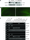
Ablation of GluRδ1 in GluRδ1−/− mice. (A) Western blot analysis of inner ear, hippocampal, and cerebellar homogenates from GluRδ1+/+ (+/+), GluRδ1+/− (+/−), and GluRδ1−/− (−/−) mice. The GluRδ1/2 antibody detected an ∼115-kDa band seen in cerebella (Cb) of GluRδ1+/+ and GluRδ1−/− mice (representing GluRδ2) and hippocampi and inner ears of GluRδ1+/+ and GluRδ1+/− mice (representing GluRδ1) but did not detect any band in GluRδ1−/− mice. Anti-β-actin antibody was used as control. (B) Immunofluorescence of hippocampal sections of a GluRδ1+/+ mouse and a GluRδ1−/− mouse at 2 months of age. The GluRδ1-specific antibody detected signals in hippocampal neurons in the GluRδ1+/+ mouse, but not in the GluRδ1−/− mouse. The bar (50 μm) applies to both panels. (C) RT-PCR analysis of different laser-captured cells from the mouse cochlear sections of GluRδ1+/+ and GluRδ1−/− mice. Lane 1, positive control cDNA of P9 whole cochlea with reverse transcription. Lane 2, negative control cDNA of P9 whole cochlea without reverse transcription. Lane 3, cDNA of all cells from the entire cochlear section. Lanes 4 to 14, cDNAs of laser-captured individual cell types. Genotypes of mice are labeled on the left. Genes for specific cell types used for RT-PCR are labeled on the right. Primers for GluRδ1 were from the deleted exons.
FIG. 3.
FIG. 3.
Cochlear thresholds are elevated at high frequencies in GluRδ1−/− (−/−) mice (A and B), and the EP is reduced in the basal turn (C). GluRδ1+/− (+/−) animals show intermediate values by all measures. Panels A and B show ABR and DPOAE data, respectively, for the same cohort of animals; panel C is from a separate group. Means and standard errors (error bars) are shown; numbers of animals (“n”) in each group are given. Statistical analyses are described in the text. Arrows on DPOAE points from GluRδ1−/− mice indicate that these values are minimum estimates because some ears showed no response at the highest sound levels (80 dB SPL). The double asterisks in panel C represent a significant difference (P was <0.01 by Student's t test; P was <0.05 by one-way ANOVA) between GluRδ1+/+ and GluRδ1−/− mice, while other group comparisons showed insignificant differences.
FIG. 4.
FIG. 4.
There is a loss of type IV fibrocytes from the spiral ligaments in the high-frequency regions of ears of GluRδ1−/− mice. Panels A and C show place-matched views of the upper basal turn (∼30-kHz region) from an ear of a GluRδ1−/− mouse and an ear of a GluRδ1+/+ mouse, respectively. Arrows point to OHCs; dotted boxes show the region of the spiral ligament where type IV fibrocytes are normally found. Panels B and D enlarge these regions. The white arrow in panel D indicates the nucleus of a type IV fibrocyte in the ear of the GluRδ1+/+ mouse; the arrow in panel B shows the absence of this cell type in the ear of the GluRδ1−/− mouse, which leaves characteristic gaps in the extracellular matrix. Panels E and F show the estimated fractional survival of IHCs, OHCs, and type IV fibrocytes in an ear of a GluRδ1−/− mouse and an ear of a GluRδ1+/+ mouse, respectively, as a function of cochlear location (converted to frequency).
FIG. 5.
FIG. 5.
Immunostaining of Kv3.1b (A and B) was reduced and lost in type IV fibrocytes (arrows) but normal in other fibrocytes of spiral ligament in the basal turn. Immunostaining of Kir4.1 (C and D), Nkcc1 (E and F), and Atp1b1 (G and H) appeared unaffected in marginal cells of stria vascularis. The bar in panel B (20 μm) applies to all panels. +/+, GluRδ1+/+; −/−, GluRδ1−/−.
FIG. 6.
FIG. 6.
GluRδ1−/− (−/−) mice are more vulnerable to acoustic injury. Temporary threshold shifts were measured in both ABR (A) and DPOAE (B) 12 h after exposure to an octave band noise at 8 to 16 kHz at 89 dB SPL for 2 h. Means and standard errors (error bars) are shown; numbers of animals in each group are in the key in panel B, which applies to both panels. Threshold shifts are not shown for the two highest test frequencies because some of the GluRδ1−/− mice showed preexposure thresholds at or near the sound pressure ceiling of our functional assay. +/+, GluRδ1+/+.
FIG. 7.
FIG. 7.
There is no change in the density of efferent innervation (A to C) or in the strength of shock-evoked efferent effects (D and E) in GluRδ1−/− mice. Panel A shows SNAP25 immunostained efferent terminals in the ear of a GluRδ1+/+ mouse. The white arrow marks a terminal in the OHC area, the black-rimmed arrow shows a terminal in the IHC area. Panels B and C show the total silhouette area of immunostained terminals as a function of cochlear location (converted to frequency) in one ear from each genotype. In the OHC area (C), values are averaged over all three OHC rows. Panel D shows representative data for each genotype from the assay used to measure cochlear efferent suppression. DPOAE amplitude (evoked with f2 at 16 kHz) was measured repeatedly before, during, and after a train of shocks to the efferent bundle; efferent suppression of DPOAE amplitude (the difference between the two dashed lines) was measured. Panel E shows average values of efferent suppression at different test frequencies obtained from a cohort of animals using the assay illustrated in panel D. Means and standard errors (error bars) are shown. +/+, GluRδ1+/+; −/−, GluRδ1−/−; +/−, GluRδ1+/−.
FIG. 8.
FIG. 8.
GluRδ1−/− mice display normal vestibular sensory epithelial morphology and function. Micrographs are from the saccular macula (A and C) and the posterior canal ampulla (B and D) of mice at 2 months of age. White arrows point to hair cell bodies; black arrowheads point to hair bundles. The scale bar in panel A applies to panels A through D. (E) The retention time (mean and SEM [error bars]) in the Rotarod test did not reveal significant differences among mice of three genotypes at 2 months of age. The number of mice tested in each group is illustrated. (F) Threshold measurements (mean and SEM [error bars]) of linear VsEPs did not show significant differences among mice of three genotypes at ages of between 2 and 3 months. The stimulus amplitude was described in dB re: 1.0g/ms and ranged from −18 to + 6 dB re: 1.0g/ms, adjusted in 3-dB steps. The number of mice tested in each group is illustrated. +/+, GluRδ1+/+; −/−, GluRδ1−/−; +/−, GluRδ1+/−.
FIG. 9.
FIG. 9.
Hippocampal morphology and function appear normal in GluRδ1−/− mice. (A) Hematoxylin and eosin staining of hippocampi in GluRδ1+/+ (+/+) and GluRδ1−/− (−/−) mice at 2 months old. No obvious differences in neuronal location, number, and position were detected between the hippocampuses of GluRδ1+/+ and GluRδ1−/− mice. Synaptic transmission appeared normal in hippocampal slices of GluRδ1+/+ and GluRδ1−/− mice at 2 months old (B and C). (B) Recordings of the extracellular fEPSPs showed that the loss of GluRδ1 did not cause any significant changes in synaptic transmission over a wide range of stimulus intensities. (C) A 200-Hz tetanic stimulation enhanced the fEPSPs to 159 ± 13% (mean ± SEM [error bars]; n = 10) and 174 ± 17% (n = 15; P was >0.05 by the Kolmogorov-Smirnov test) of their initial levels, when measured 60 min after tetanization in slices from GluRδ1−/− and GluRδ1+/+ mice, respectively. (D) Performance in the place task of the water maze did not show significant differences among GluRδ1+/+, GluRδ1+/− (+/−), and GluRδ1−/− mice at 2 to 3 months of age (n = 6, 10, 11, respectively). Mean latency to reach the hidden platform is shown on each of the 10 test days. All groups performed similarly in this task. The vertical bar indicates one standard error of the difference (SED) in group means.

References

    1. Antoli-Candela, F. J., and N. Y. S. Kiang. 1978. Unit activity underlying the N1 potential, p. 165-191. In R. F. Naunton and C. Fernández (ed.), Evoked electrical activity in the auditory nervous system. Academic Press, New York, NY.
    1. Ben-Yosef, T., I. A. Belyantseva, T. L. Saunders, E. D. Hughes, K. Kawamoto, C. M. Van Itallie, L. A. Beyer, K. Halsey, D. J. Gardner, E. R. Wilcox, J. Rasmussen, J. M. Anderson, D. F. Dolan, A. Forge, Y. Raphael, S. A. Camper, and T. B. Friedman. 2003. Claudin 14 knockout mice, a model for autosomal recessive deafness DFNB29, are deaf due to cochlear hair cell degeneration. Hum. Mol. Genet. 12:2049-2061. - PubMed
    1. Boettger, T., C. A. Hubner, H. Maier, M. B. Rust, F. X. Beck, and T. J. Jentsch. 2002. Deafness and renal tubular acidosis in mice lacking the K-Cl co-transporter Kcc4. Nature 416:874-878. - PubMed
    1. Cohen-Salmon, M., T. Ott, V. Michel, J. P. Hardelin, I. Perfettini, M. Eybalin, T. Wu, D. C. Marcus, P. Wangemann, K. Willecke, and C. Petit. 2002. Targeted ablation of connexin26 in the inner ear epithelial gap junction network causes hearing impairment and cell death. Curr. Biol. 12:1106-1111. - PMC - PubMed
    1. Darrow, K. N., S. F. Maison, and M. C. Liberman. 2007. Selective removal of lateral olivocochlear efferents increases vulnerability to acute acoustic injury. J. Neurophysiol. 97:1775-1785. - PMC - PubMed

Publication types

MeSH terms

Substances

LinkOut - more resources