Review
. 2007 May;3(5):252-62.
doi: 10.1038/nchembio876.

Foldamers as versatile frameworks for the design and evolution of function

Affiliations
Review

Foldamers as versatile frameworks for the design and evolution of function

Catherine M Goodman et al. Nat Chem Biol. 2007 May.

Abstract

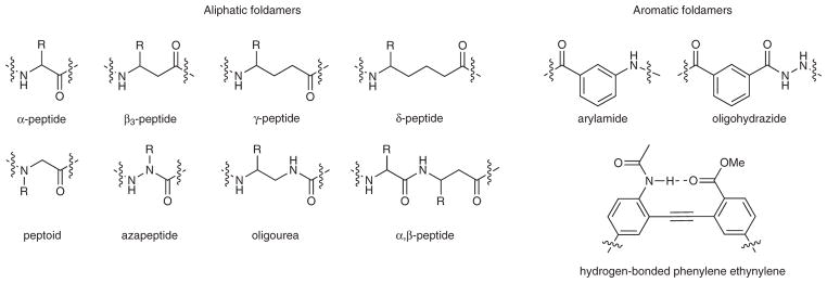
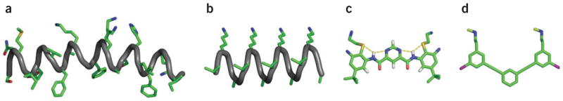
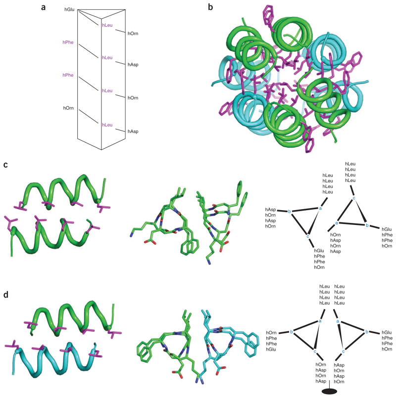
Foldamers are sequence-specific oligomers akin to peptides, proteins and oligonucleotides that fold into well-defined three-dimensional structures. They offer the chemical biologist a broad pallet of building blocks for the construction of molecules that test and extend our understanding of protein folding and function. Foldamers also provide templates for presenting complex arrays of functional groups in virtually unlimited geometrical patterns, thereby presenting attractive opportunities for the design of molecules that bind in a sequence- and structure-specific manner to oligosaccharides, nucleic acids, membranes and proteins. We summarize recent advances and highlight the future applications and challenges of this rapidly expanding field.


Conflict of interest statement

COMPETING INTERESTS STATEMENT

The authors declare competing financial interests: details accompany the full-text HTML version of the paper at www.nature.com/naturechemicalbiology/.

Figures

Figure 1
Figure 1
Examples of foldamer frameworks.
Figure 2
Figure 2
The 14-helix conformation. (a) The most studied β-peptide helix, the 14-helix, is named as such because of the 14-membered ring formed when the i and i + 3 amides form a hydrogen bond. (b) Crystal structure of a 14-helix highlighting backbone hydrogen bonds. (c) 14-helix conformation depicting N-terminal and C-terminal capping motifs. Also shown, electrostatic pairing between oppositely charged side chains at i and i + 3 residues, as well as hydrophobic side chains at i, i + 3 that are clustered in the crystal structure. Crystal structures are from ref. .
Figure 3
Figure 3
14-helix and mixed α/β-helices. Interactions between substituents at C3 of residues i, i + 3 in the 14-helix; distances range from 4.7 Å to 5.2 Å in the crystal structure of Zwit-1F (ref. 61). The corresponding cross-strand distance ran1ges from 4.5 Å to 5.5 Å in a β-sheet (Protein Data Bank (PDB) code 1TTA), and the i, i + 4 distances range from 5.9 Å to 6.5 Å in an α-helix (PDB code 2ZTA). Mixed α/β-peptide (side and top views shown): overlay of the α-helix and a helix formed in a mixed α/β-peptide (the additional backbone C2 methylene groups are shown as spheres in the β-amino acid) (PDB codes 2ZTA and 2OXJ).
Figure 4
Figure 4
Design principles for foldamer scaffolds. (ac) Examples of hydrogen bonding patterns used in foldamer scaffolds that have been shown to form helix-like structures with indicated repeating subunits. (d) Linear foldamer stabilized by hydrogen bonding network. (e) The precise spacing of carboxylates on an arylamide framework promotes the formation of calcite (CaCO3) crystals in a specific morphology.
Figure 5
Figure 5
Self-associating foldamer. Structure and association of monomeric and dimeric foldamers.
Figure 6
Figure 6
Downsizing a natural host defense peptide. (a) The natural host defense peptide magainin (PDB code 2MAG). (b) 14-helix antimicrobial β-peptide. (c) Arylamide antimicrobial foldamer. (d) Phenylalkylnyl-based antimicrobial compound.
Figure 7
Figure 7
Simultaneous parallel and antiparallel hydrophobic packing. Crystal structure of designed β-peptide is shown; parallel dimer pairs are in blue and green. (a) β-peptide sequence mapped onto helical wheel. (b) Full octomeric structure, highlighting the parallel monomer chains within each subunit (hydrophobic packing of leucine and phenylalanine shown in pink). (c) Parallel helical interface with leucine packing highlighted. Also shown: top view, helical wheel. (d) Antiparallel helical interface with leucine packing highlighted. Also shown: top view, helical wheel.

References

    1. DeGrado WF, Summa CM, Pavone V, Nastri F, Lombardi A. De novo design and structural characterization of proteins and metalloproteins. Annu Rev Biochem. 1999;68:779–819. - PubMed
    1. Baker D. Prediction and design of macromolecular structures and interactions. Phil Trans R Soc Lond B. 2006;361:459–463. - PMC - PubMed
    1. Alvizo O, Allen BD, Mayo SL. Computational protein design promises to revolutionize protein engineering. Biotechniques. 2007;42:31, 33, 35. passim. - PubMed
    1. Gellman SH. Foldamers: a manifesto. Acc Chem Res. 1998;31:173–180.
    1. Rueping M, Mahajan Y, Sauer M, Seebach D. Cellular uptake studies with β-peptides. Chem Bio Chem. 2002;3:257–259. - PubMed