

Role of a novel excipient poly(ethylene glycol)-b-poly(L-histidine) in retention of physical stability of insulin at aqueous/organic interface
- PMID: 17439239
- PMCID: PMC2562025
- DOI: 10.1021/mp060120z
Role of a novel excipient poly(ethylene glycol)-b-poly(L-histidine) in retention of physical stability of insulin at aqueous/organic interface
Abstract
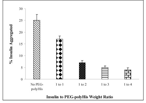
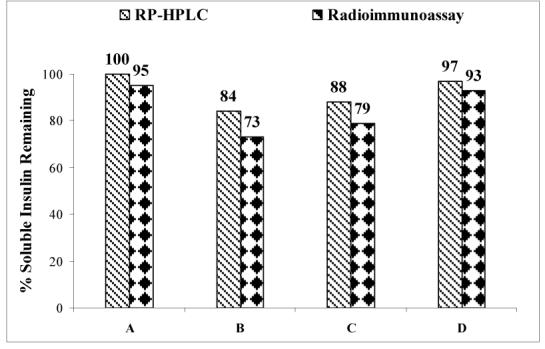
The aim of this study was to investigate whether a cationic polyelectrolyte, poly(ethylene glycol)-b-poly(L-histidine) diblock copolymer (PEG-polyHis), can stabilize insulin, at the aqueous/methylene chloride interface formed during the microencapsulation process. Insulin aggregation at this interface was monitored spectrophotometrically at 276 nm. The effects of protein concentration, pH of the aqueous medium, and the presence of poly(lactic-co-glycolic acid) (PLGA) in methylene chloride (MC) on insulin aggregation were observed. For the 2.0 mg/mL insulin solutions in phosphate buffer (PB), the effect of addition of Pluronic F-127 as a positive control and addition of PEG-polyHis as a novel excipient in PB was also evaluated at various insulin/polymeric excipient weight ratios. The conformation of insulin protected by PEG-polyHis and recovered after interfacial exposure was evaluated via circular dichroism (CD) spectroscopy. Greater loss in soluble insulin was observed with increasing insulin concentrations. pH 6.0 was selected for optimal ionic interactions between insulin and PEG-polyHis based on zeta potential and particle size studies. pH 4.5 and 7.4 (no ionic complexation between insulin and PEG-polyHis) were selected as controls to compare the stabilization effect of PEG-polyHis with that at pH 6.0. Incubation of PEG-polyHis with insulin at pH 6.0 drastically reduced protein aggregation, even in the presence of PLGA. PEG-polyHis and F-127 reduced insulin aggregation in noncomplexing pH conditions pointing to the role played by PEG in modulation of insulin adsorption at the interface. Far-UV (205-250 nm) CD study revealed negligible qualitative effects on soluble insulin's secondary structure after interfacial exposure. RP-HPLC and size-exclusion HPLC showed no deamidation of insulin or formation of soluble high molecular weight transformation products respectively. MALDI-TOF mass spectrometry confirmed the results from chromatographic procedures. Radioimmunoassay carried out on select samples showed that recovered soluble insulin had retained its immunoreactivity. An experimental method to simulate interfacial denaturation of proteins was designed for assessment of protein stability at the interface and screening for novel protein stabilizers. Understanding and manipulation of such polyelectrolyte-insulin complexation will likely play a role in insulin controlled delivery via microsphere formulation(s).
Figures


References
-
- Varde NK, Pack DW. Microspheres for controlled release drug delivery. Expert Opin. Biol. Ther. 2004;4(1):35–51. - PubMed
-
- Wise DL, Trantolo DJ, Marino RT, Kitchell JP. Opportunites and challenges in the design of implantable biodegradable polymeric systems for the delivery of aitimicrobial agents and vaccines. Adv. Drug Delivery Rev. 1987;1:19–39.
-
- Cohen S, Yoshioka T, Lucarelli M, Hwang LH, Langer R. Controlled delivery systems for proteins based on poly(lactic/glycolic acid) microspheres. Pharmaceutical Research. 1991;8:713–720. - PubMed
-
- Wang YJ, Hanson M. Parenteral formulations of peptides and proteins: Stability and stabilizers. J. Parenteral Sci. Tech. 1988;42:1–26.
-
- van de Weert M, Hoechstetter J, Hennink WE, Crommelin DJ. The effect of a water/organic solvent interface on the structural stability of lysozyme. J. Control. Release. 2000;68:351–359. - PubMed
Publication types
MeSH terms
Substances
Grants and funding
LinkOut - more resources
Full Text Sources
Other Literature Sources
Medical
