. 2007 May;5(5):e102.
doi: 10.1371/journal.pbio.0050102.

High incidence of non-random template strand segregation and asymmetric fate determination in dividing stem cells and their progeny

Affiliations

High incidence of non-random template strand segregation and asymmetric fate determination in dividing stem cells and their progeny

Michael J Conboy et al. PLoS Biol. 2007 May.

Erratum in

  • PLoS Biol. 2007 Jul;5(7):e182

Abstract

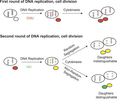
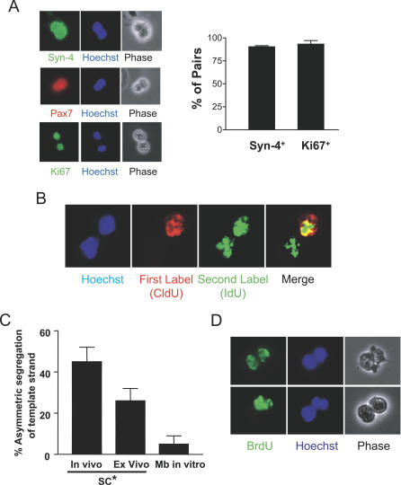
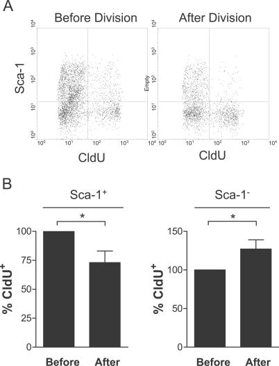
Decades ago, the "immortal strand hypothesis" was proposed as a means by which stem cells might limit acquiring mutations that could give rise to cancer, while continuing to proliferate for the life of an organism. Originally based on observations in embryonic cells, and later studied in terms of stem cell self-renewal, this hypothesis has remained largely unaccepted because of few additional reports, the rarity of the cells displaying template strand segregation, and alternative interpretations of experiments involving single labels or different types of labels to follow template strands. Using sequential pulses of halogenated thymidine analogs (bromodeoxyuridine [BrdU], chlorodeoxyuridine [CldU], and iododeoxyuridine [IdU]), and analyzing stem cell progeny during induced regeneration in vivo, we observed extraordinarily high frequencies of segregation of older and younger template strands during a period of proliferative expansion of muscle stem cells. Furthermore, template strand co-segregation was strongly associated with asymmetric cell divisions yielding daughters with divergent fates. Daughter cells inheriting the older templates retained the more immature phenotype, whereas daughters inheriting the newer templates acquired a more differentiated phenotype. These data provide compelling evidence of template strand co-segregation based on template age and associated with cell fate determination, suggest that template strand age is monitored during stem cell lineage progression, and raise important caveats for the interpretation of label-retaining cells.


Conflict of interest statement

Competing interests. The authors have declared that no competing interests exist.

Figures

Figure 1
Figure 1. Models of Template Strand Segregation during Cell Division
Models of random versus non-random segregation of template strand DNA. Only two pairs of chromosomes are shown for clarity. Newly synthesized DNA strands are indicated by color and dashed lines. At the first division, the newly synthesized DNA is labeled with CldU, is complementary to the template DNA, and thus is inherited equally. For the next round of DNA synthesis (during which the newly synthesized strand is labeled with IdU), one template strand is the older, unlabeled strand, and the other is the younger, CldU-labeled strand. If the cell segregates the chromatids with the older templates (and their complementary IdU-labeled strands) to one daughter, then all of the chromatids containing the younger, CldU-label templates would be inherited by the other daughter (also along with their complementary IdU-labeled strands). Note that this asymmetric inheritance using pulsed labels is only detectable after the second division when the strands labeled during a previous round of DNA replication have become templates for a subsequent round of DNA replication and inheritance.
Figure 2
Figure 2. Evidence of Co-Segregation of DNA Template Strands during Muscle Progenitor Cell Division
(A) Following muscle injury, CldU was administered to coincide with an early round of satellite cell division (~day 2 after injury), and IdU was administered to coincide with the subsequent round of DNA replication in the population. Cells were then isolated from muscle and plated singly on coated chamber slides, allowed to complete the current cell division, and then fixed. Cell pairs were analyzed for the expression of Syndecan-4 and Pax7, myogenic lineage markers [9,27], and for the marker of cell proliferation, Ki67. Representative pairs are shown on the left. The percentages of pairs that were myogenic (Syndecan-4+) and proliferating (Ki67+) were quantified and are presented graphically on the right. (B) Cell pairs were immunostained for CldU and IdU. Shown is a representative photograph of an immunostained pair of cells, in which both daughter cells were labeled with the second label, IdU (green), but only one daughter inherited the first label, CldU (red). (C) Quantification of asymmetric pairs. Almost half of the pairs derived from cells isolated from regenerating muscle showed asymmetric CldU staining as in (A) (all had symmetric IdU staining), and almost one third of the pairs of satellite cell progeny activated ex vivo in bulk myofiber explant cultures showed asymmetric inheritance of DNA label. Only a small percentage of pairs derived from established myoblast cultures (Mb in vitro) showed asymmetric inheritance of DNA label. Data represent mean ± the standard deviation (SD) (n = 4–7). SC*, progeny of activated satellite cells. (D) Muscle was injured and labeled with BrdU on day 2 of regeneration, as with CldU above (C). On day 3, the satellite cell progeny were prepared, plated sparsely, and cytochalasin D was added to arrest cytokinesis. The top row shows a cell pair symmetrically inheriting BrdU-labeled DNA, and the bottom row shows an asymmetric pair. Similar percentages of cells showed asymmetric inheritance of template strands using this protocol as in (C).
Figure 3
Figure 3. Divergent Cell Fates Associated with Asymmetric Segregation of Template Strands
(A) Pairs of cells as in Figure 2 were co-immunostained for BrdU and the myoblast marker Desmin; representative images are shown. (B) The data from experiments as in (A) were quantified. Pairs with Desmin expression and asymmetric BrdU labeling were distinguished based on the pattern of Desmin expression (asymmetric and coincident with BrdU, asymmetric and mutually exclusive with BrdU, or symmetric). The legend for individual cells is shown to the right, and the legends for the cell pairs are shown below. Data represent mean ± SEM (n = 4).
Figure 4
Figure 4. Segregation of the Oldest Template Strands to the Less Differentiated Progeny
(A) Cells pairs were co-immunostained for Sca-1 and Desmin. Shown is a representative pair of cells demonstrating Sca-1 expression in one daughter and Desmin expression in the other. (B) The data from experiments as in (A) were quantified. Pairs with Sca-1 expression and asymmetric Desmin staining were distinguished based on the pattern of Sca-1 expression (asymmetric and mutually exclusive with Desmin, asymmetric and coincident with Desmin, or symmetric). The legends for the cells are as described for Figure 3B. Data represent mean ± S.E.M. (n = 4).
Figure 5
Figure 5. Correlation of Template Strand Segregation with Cell Fates in Myogenic Progenitors
Cells were labeled and isolated from regenerating muscle as in Figure 2. Half of the cells were fixed immediately (Before Division). The other half (After Division) was cultured overnight to allow the cells to complete the current cell cycle and divide. The cells were then harvested and analyzed by FACS for Sca-1, the labeled (younger) template (CldU), and newly synthesized DNA (IdU). Gatings were on forward scatter (FSC) and IdU-labeled cells. (A) A representative FACS plot of Sca-1 versus CldU is shown in the population before and after cell division. (B) Quantitation of FACS plots as shown in (A) demonstrates the decrease in the percentage of Sca-1+ cells that have labeled template strands before and after cell division and the corresponding increase in the percentage of Sca-1 cells having labeled template strands (n ≥ 3; a single asterisk [*] indicates p < 0.01).

Comment in

References

    1. Cairns J. Mutation selection and the natural history of cancer. Nature. 1975;255:197–200. - PubMed
    1. Lark KG, Consigli RA, Minocha HC. Segregation of sister chromatids in mammalian cells. Science. 1966;154:1202–1205. - PubMed
    1. Potten CS, Hume WJ, Reid P, Cairns J. The segregation of DNA in epithelial stem cells. Cell. 1978;15:899–906. - PubMed
    1. Merok JR, Lansita JA, Tunstead JR, Sherley JL. Cosegregation of chromosomes containing immortal DNA strands in cells that cycle with asymmetric stem cell kinetics. Cancer Res. 2002;62:6791–6795. - PubMed
    1. Karpowicz P, Morshead C, Kam A, Jervis E, Ramunas J, et al. Support for the immortal strand hypothesis: Neural stem cells partition DNA asymmetrically in vitro. J Cell Biol. 2005;170:721–732. - PMC - PubMed

Publication types