. 2007 Apr;24(4):657-73.
doi: 10.1089/neu.2006.0011.

Electromagnetic controlled cortical impact device for precise, graded experimental traumatic brain injury

Affiliations

Electromagnetic controlled cortical impact device for precise, graded experimental traumatic brain injury

David L Brody et al. J Neurotrauma. 2007 Apr.

Abstract

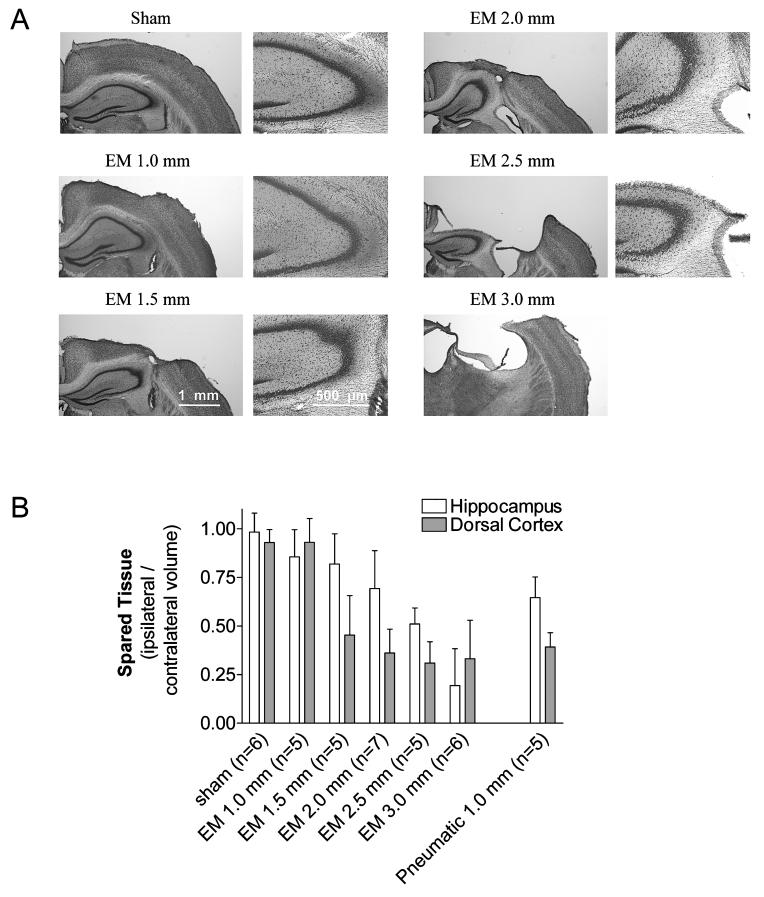
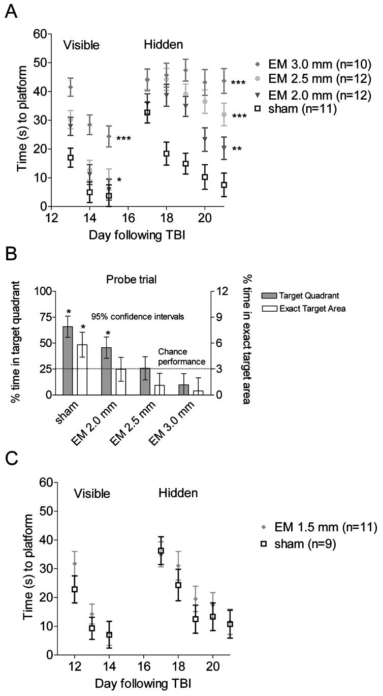
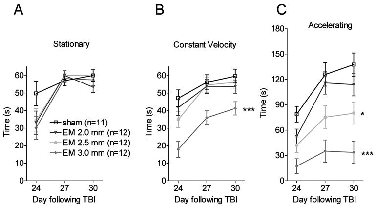
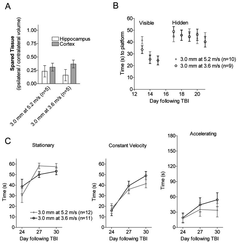
Genetically modified mice represent useful tools for traumatic brain injury (TBI) research and attractive preclinical models for the development of novel therapeutics. Experimental methods that minimize the number of mice needed may increase the pace of discovery. With this in mind, we developed and characterized a prototype electromagnetic (EM) controlled cortical impact device along with refined surgical and behavioral testing techniques. By varying the depth of impact between 1.0 and 3.0 mm, we found that the EM device was capable of producing a broad range of injury severities. Histologically, 2.0-mm impact depth injuries produced by the EM device were similar to 1.0-mm impact depth injuries produced by a commercially available pneumatic device. Behaviorally, 2.0-, 2.5-, and 3.0-mm impacts impaired hidden platform and probe trial water maze performance, whereas 1.5-mm impacts did not. Rotorod and visible platform water maze deficits were also found following 2.5- and 3.0-mm impacts. No impairment of conditioned fear performance was detected. No differences were found between sexes of mice. Inter-operator reliability was very good. Behaviorally, we found that we could statistically distinguish between injury depths differing by 0.5 mm using 12 mice per group and between injury depths differing by 1.0 mm with 7-8 mice per group. Thus, the EM impactor and refined surgical and behavioral testing techniques may offer a reliable and convenient framework for preclinical TBI research involving mice.


Figures

FIG. 1
FIG. 1
Design of an electromagnetic controlled cortical impact device for experimental traumatic brain injury. (A) Photograph of the impactor device mounted on the left arm of a stereotaxic device. Motorized drill with 5-mm trephine mounted on the right arm. (B) Schematic of the components of the impact system. Control signals from a Windows-based notebook computer running custom Matlab™ routines are fed through a digital-to-analog converter. The digital-to-analog converter output is sent to a servo amplifier. The servo amplifier transmits current from 72-V power supply to the impactor containing an electromagnetic voice coil. This voice coil drives the impactor. (C) Photograph of the impactor tip in the raised position. A laser-Doppler displacement sensor was used to measure velocity of the tip during the impact stroke.
FIG. 2
FIG. 2
Trajectories of the impactor tip during impact stroke: measurements of velocity and overshoot. All trajectories were measured using a fast laser Doppler displacement sensor aimed at the tip of the impactor. (A) EM impactor set at 3 or 7 V, yielding velocities of 3.6 or 5.2 m/sec. Overshoot, defined as the transient excursion of the impactor tip past the set distance specified by the user, was 0.31 ± 0.032 mm at 7 V and essentially unchanged at 3 V. Set distance and overshoot are indicated by top and bottom dashed lines, respectively. (B) Pneumatic impactor (Amscien, AMS201) with high-pressure settings of 50 or 100 psi yielding velocities of 3.7 or 5.2 m/sec. (C) Overshoot as a function of velocity for the electromagnetic and pneumatic impactors. Overshoot was strongly velocity-dependent for the pneumatic impactor as tested but there was little change in overshoot with velocity for the electromagnetic impactor.
FIG. 3
FIG. 3
Histological analysis of injuries produced by EM and pneumatic devices. (A) Histological images of cresyl violet-stained coronal sections following sham injury, and 1.0, 1.5, 2.0, 2.5, and 3.0 mm impact using the EM device. Left images in each pair show overall dorsal cortical and hippocampal tissue loss and architecture. Right image in each pair show further details of hippocampal architecture. Cortical and hippocampal tissue injury and anatomical distortion increase in a graded fashion with increased impact depth. All images were obtained from slices through the same anatomical region (bregma -1.7 mm), selected based on the architecture of the contralateral hemisphere. (B) Proportion of spared ipsilateral hippocampus and dorsal cortex remaining 1 month after TBI. Each stereologically determined hippocampal or cortical volume was normalized by the corresponding contralateral volume for that animal. Set depth of injury on the x axis. Error bars represent standard deviations. All injuries were performed by the same investigator and volumes were assessed without knowledge of device or impact set depth.
FIG. 4
FIG. 4
Behavioral characterization of mice injured using the EM device in the Morris water maze. (A) Time to reach the platform as a function of day following experimental TBI. During visible platform testing, two of 12 mice subjected to 3.0-mm impacts failed to reliably swim to the platform and were disqualified from further water maze testing. No mice in the sham, 2.0-mm, or 2.5-mm impact groups were disqualified. Data shown represent mean and standard errors from the remaining, non-disqualified mice. During hidden platform testing, thre was a clear gradation in performance, with more severely injured animals performing worse. (***p = 0.0002, **p 0.006, *p = 0.039, repeated-measures ANOVA followed by Tukey HSD posthoc test for comparisons vs. sham). (B) Water maze probe testing; the platform was removed after the last day of hidden platform testing and mice were placed in the pool for a single, 30-sec trial. Sham mice spent considerably more time in the target quadrant and exact area where the platform had been than would have been expected by chance. Mice subjected to TBI had impaired performance, again in a graded fashion depending on the severity of injury (*95% confidence interval did not overlap with the performance expected by chance.) (C) In a separate experiment, mice subjected to a 1.5-mm impact did not show significant water maze performance deficits compared with concurrently tested, sham-injured mice.
FIG. 5
FIG. 5
Rotorod performance of mice injured using the EM device. Each mouse was tested on three separate days with one 60-sec stationary rod trial, two 60-sec constant velocity trials, and two 3-min accelerating trials. Data shown represent means and standard errors. All mice were included, even those disqualified from water maze testing. (A) Stationary rod performance. All mice performed well, even those subjected to the most severe injuries. (B) Constant velocity performance. Mice subjected to 3.0-mm impacts had impaired abilities to stay on the rod (***p = 0.0002 vs. sham) whereas milder injuries did not disrupt this ability. (C) Accelerating rod performance. There was a gradation of impairment with more severely injured animals performing worse (*p = 0.016, ***p = 0.0002 vs. sham).
FIG. 6
FIG. 6
Statistical power to distinguish between two injury severities in terms of hidden platform water maze performance. Monte-Carlo simulations were used based on original data sets from the 22 mice in the 2.0-mm and 3.0-mm impact groups. 150 random subsamples of these 22 mice were reanalyzed using repeated-measures ANOVA. For this analysis, the total number of mice in the random subsamples is plotted on the x-axis. The median and 80% confidence interval of the resulting p-values are plotted on the y-axis. This analysis indicates that, with 15 or greater total mice in the two groups, we would expect at least an 80% likelihood of detecting a difference between groups with a p-value of <0.05.
FIG. 7
FIG. 7
Injury severity as a function of impact velocity. An additional group of WT mice were injured at 3.0-mm impact depth at a velocity of 3.6 m/sec and compared with 3.0-mm impats produced at a velocity of 5.2 m/sec. Data from the 5.2 m/sec groups is the same as that shown in Figures 3-5. (A) Fraction of spared tissue in hippocampus and cortex, assessed histologically, was not significantly different between groups. (B) Morris water maze performance. Two of 11 mice in the 3.0-mm at 3.6 m/sec group were disqualified from Morris water maze testing because of poor visible platform performance. The behavioral deficits in both visible and hidden platform performance were equally severe in both groups. (C) Rotored performance deficits were similar in the two groups.

References

    1. ABRAHAMSON EE, IKONOMOVIC MD, CIALLELLA JR, et al. Caspase inhibition therapy abolishes brain trauma-induced increases in Abeta peptide: implications for clinical outcome. Exp. Neurol. 2006;197:437–450. - PubMed
    1. BAYIR H, CLARK RS, KOCHANEK PM. Promising strategies to minimize secondary brain injury after head trauma. Crit. Care Med. 2003;31:S112–S117. - PubMed
    1. BAYIR H, KAGAN VE, BORISENKO GG, et al. Enhanced oxidative stress in iNOS-deficient mice after traumatic brain injury: support for a neuroprotective role of iNOS. J. Cereb. Blood Flow Metab. 2005;25:673–684. - PubMed
    1. BAYLY PV, DIKRANIAN KT, BLACK EE, et al. Spatiotemporal evolution of apoptotic neurodegeneration following traumatic injury to the developing rat brain. Brain Res. 2006;1107:70–81. - PMC - PubMed
    1. BERMPOHL D, YOU Z, KORSMEYER SJ, MOSKOWITZ MA, WHALEN MJ. Traumatic brain injury in mice deficient in Bid: effects on histopathology and functional outcome. J. Cereb. Blood Flow Metab. 2006;26:625–633. - PubMed

Publication types

LinkOut - more resources