. 2007 Apr 17:7:33.
doi: 10.1186/1471-213X-7-33.

Identification of two novel glial-restricted cell populations in the embryonic telencephalon arising from unique origins

Affiliations

Identification of two novel glial-restricted cell populations in the embryonic telencephalon arising from unique origins

Frederick G Strathmann et al. BMC Dev Biol. .

Abstract

Background: Considerably less attention has been given to understanding the cellular components of gliogenesis in the telencephalon when compared to neuronogenesis, despite the necessity of normal glial cell formation for neurological function. Early proposals of exclusive ventral oligodendrocyte precursor cell (OPC) generation have been challenged recently with studies revealing the potential of the dorsal telencephalon to also generate oligodendrocytes. The identification of OPCs generated from multiple regions of the developing telencephalon, together with the need of the embryonic telencephalon to provide precursor cells for oligodendrocytes as well as astrocytes in ventral and dorsal areas, raises questions concerning the identity of the precursor cell populations capable of generating macroglial subtypes during multiple developmental windows and in differing locations.

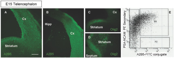
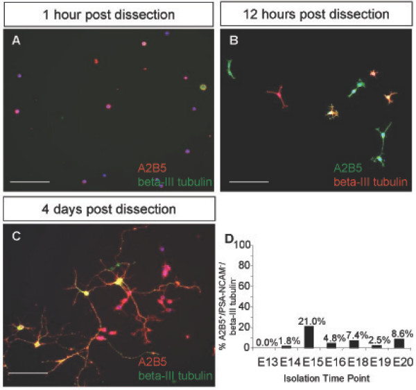
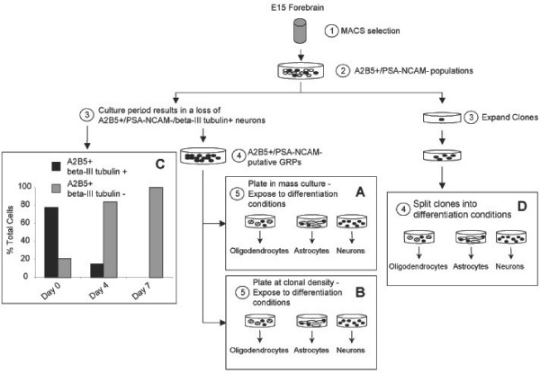
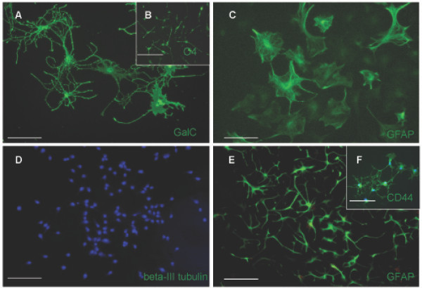
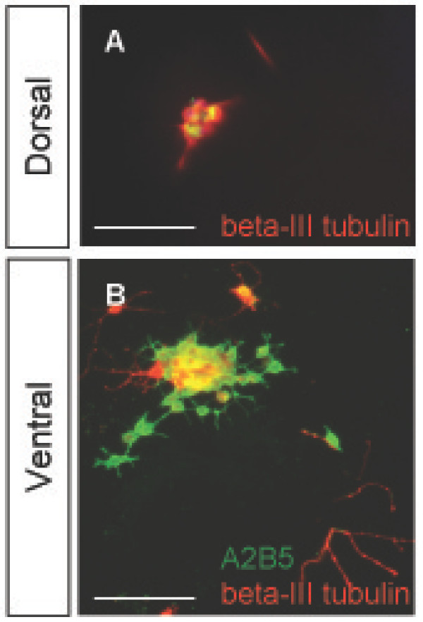
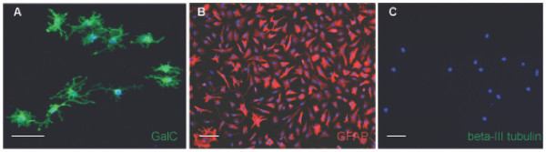
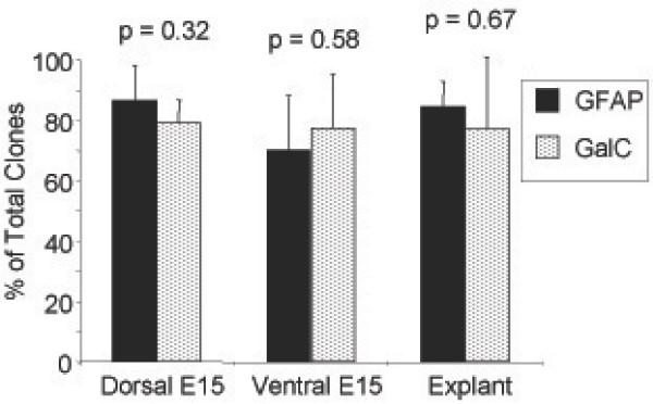
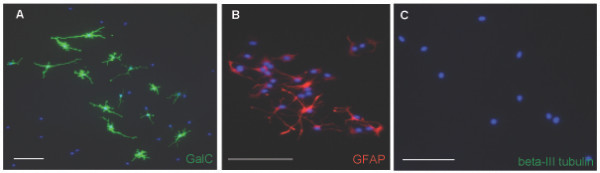
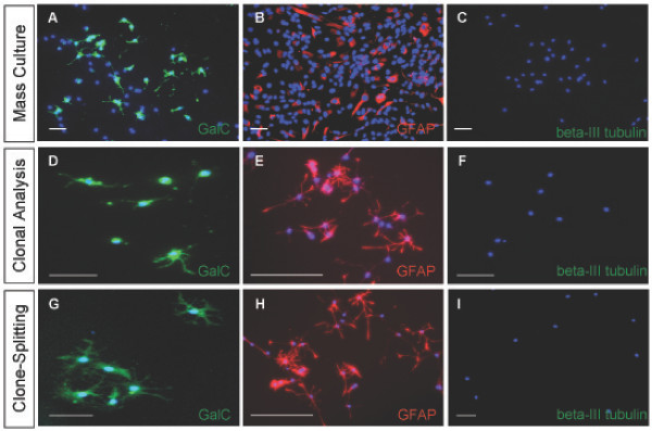
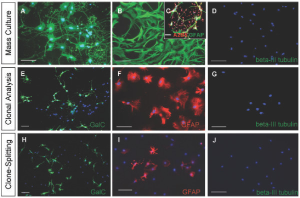
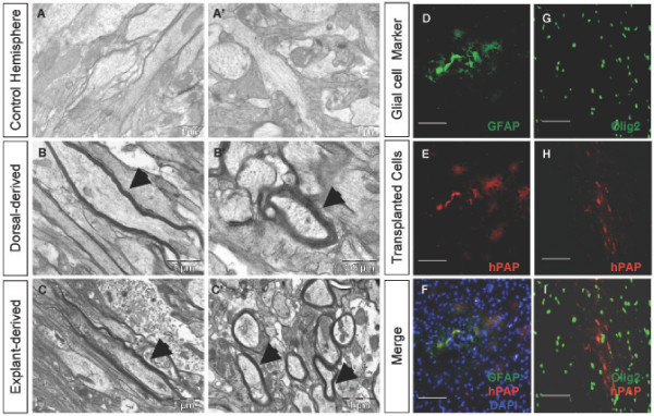
Results: We have identified progenitor populations in the ventral and dorsal telencephalon restricted to the generation of astrocytes and oligodendrocytes. We further demonstrate that the dorsal glial progenitor cells can be generated de novo from the dorsal telencephalon and we demonstrate their capacity for in vivo production of both myelin-forming oligodendrocytes and astrocytes upon transplantation.

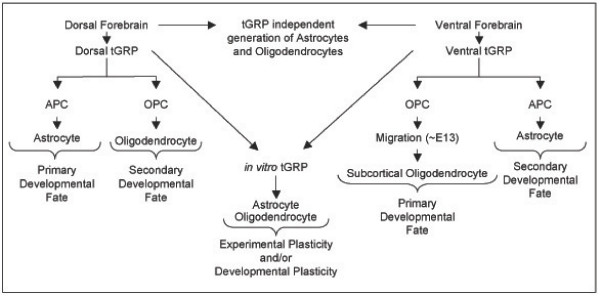
Conclusion: Based on our results we offer a unifying model of telencephalic gliogenesis, with the generation of both oligodendrocytes and astrocytes from spatially separate, but functionally similar, glial restricted populations at different developmental times in the dorsal and ventral CNS.


Figures

Figure 1
Figure 1
A2B5+ cells in the telencephalon. (A), A2B5+ cells are seen in coronal sections of the developing striatum and dorsolateral neocortex of the E15 telencephalon. (B) A2B5+ cells are absent in the developing hippocampal region. (C,D) The dorsal A2B5+ region is not Olig2+ (C) while the ventral A2B5+ region partially overlaps with the Olig2+ domain in the developing striatum (D). (E) FACS data of A2B5+/PSA-NCAM- stained cells shows three cell populations, including PSA-NCAM+, A2B5+/PSA-NCAM+, and A2B5+. Scale bar, 100 μm.
Figure 2
Figure 2
A subset of A2B5+ cells are also beta-III tubulin+ in the E15 dorsal telencephalon. (A-C) The isolated A2B5+/PSA-NCAM- cell population from the dorsal telencephalon included a beta-III tubulin+ population, seen at 1 hour (A), 12 hours (B), and 4 days (C) post isolation. (D) Isolated A2B5+/PSA-NCAM- cells were stained and analyzed for beta-III tubulin presence between E13 and E20. E15 was determined to be the peak time to isolate A2B5+/PSA-NCAM-/beta-III tubulin- cells as 22% of the E15 A2B5+/PSA-NCAM- population was beta-III tubulin-. DAPI, blue nuclear stain. Scale bars, 100 μm.
Figure 3
Figure 3
Outline of the isolation procedure used to characterize the putative glial restricted progenitor population. A2B5+/PSA-NCAM- cells were selected by MACS resulting in a heterogeneous mixture of cells. For mass culture studies (A) and clonal analysis (B), cells were maintained in culture for two cell passages to select for proliferative cells and remove the A2B5+ neuronal population (C). The resultant putative glial restricted progenitor population was then plated at mass culture or clonal density and exposed to differentiating conditions including a pro-oligodendrocytic condition, a pro-astrocytic condition, or a pro-neuronal condition (see Materials and Methods). Alternatively, the heterogeneous mixture of cells obtained from the MACS selection was plated at clonal density, and resultant clones were selectively passaged and split into the differentiation conditions (D).
Figure 4
Figure 4
The putative dorsal glial restricted progenitor population can generate macroglial subtypes in mass culture. (A,C,D) Putative glial restricted progenitor cells generate GalC+ cells (A) GFAP+ cells (C) but do not generate neurons (D) after 6 days of exposure to the appropriate differentiation conditions (see Results). (B) After 4 days of growth in the pro-oligodendrocyte condition, O4+ cells were readily identifiable. (E,F) Exposure of the putative glial restricted progenitor population to BMP-4 is insufficient to result in detection of the known astrocyte marker GFAP until 10 days (E), but does induce the Astrocyte Precursor Cell marker, CD44, after 6 days (F). DAPI, blue nuclear stain (D,F). Scale bars, 100 μm.
Figure 5
Figure 5
Neuron generation from E15 unsorted dorsal and ventral telencephalic cells. In order to validate the pro-neuronal condition used, cells present in the E15 dorsal (A) and ventral (B) telencephalon before MACS selection were exposed to the pro-neuronal condition used for glial restricted progenitor characterization and were found to generate beta-III tubulin+ cells after 6 days in culture. Scale bars, 100 μm.
Figure 6
Figure 6
Clonal analysis of the putative dorsal glial restricted progenitor further indicates glial restriction. (A-C) To distinguish between the potential presence of an APC/OPC cell mixture and the presence of a glial restricted progenitor population, the putative glial restricted progenitor population was grown at clonal density and exposed to the differentiating conditions, resulting in the detection of clones containing GalC+ cells (A) clones containing GFAP+ cells (B) but no neuron containing clones (C). DAPI blue nuclear stain. Scale bars, 100 μm.
Figure 7
Figure 7
Clonal Summary. A summary of the generated clones from Dorsal, Ventral, and Explant derived glial restricted progenitor is provided, with no significant difference (p > 0.05; Student's t-test) between astrocyte and oligodendrocyte containing clone numbers.
Figure 8
Figure 8
Clone splitting confirms the ability of the putative glial restricted progenitor cell to generate both oligodendrocytes and astrocytes. Split clones (see Results) of A2B5+/PSA-NCAM- founder cells can generate GalC+ cells (A) GFAP+ cells (B) but not neurons (C) and allows for the classification of the A2B5+/PSA-NCAM-/beta-III tubulin- cell as a glial restricted progenitor cell. DAPI, blue nuclear stain. Scale bars, 100 μm.
Figure 9
Figure 9
The dorsal telencephalon has the potential to generate glial restricted progenitor cells independent of ventral cell infiltration. (A-C) Cells with the similar antigenic profile described for the dorsal glial restricted progenitor population were isolated from two day in vitro grown dorsal explants, and can generate GalC+ cells (A) GFAP+ cells (B) but not neurons (C) in mass culture. (D-F) Explant derived putative glial restricted progenitors can generate clones containing GalC+ cells (D) clones containing GFAP+ cells (E) but no clones containing neurons (F) when exposed to the differentiation conditions. (G-I) Split clones of explant derived putative glial restricted progenitor founder cells can generate GalC+ cells (G) GFAP+ cells (H) but not neurons (I). DAPI, blue nuclear stain. Scale bars, 100 μm.
Figure 10
Figure 10
A glial restricted progenitor population cell can be isolated from the E15 ventral telencephalon. (A,B,D) Putative glial restricted progenitor cells sharing the similar antigenic profile of the dorsal glial restricted progenitor population were isolated from the E15 ventral telencephalon, consisting of the AEP and MGE. This cell population generated GalC+ cells (A) GFAP+ cells (B) but not neurons (D) in mass culture. (C) Putative glial restricted progenitor cells do not make A2B5+/GFAP+ type-2 astrocytes in response to CNTF. (E-G), To distinguish between APC/OPC presence and glial restricted progenitor presence, ventral putative glial restricted progenitor cells were grown at clonal density and generated GalC+ cells (E) GFAP+ cells (F) but not neurons (G) when examined at the clonal level. (H-J) Split clones of ventral putative glial restricted progenitor founder cells generated GalC+ cells (H) GFAP+ cells (I) but not neurons (J). DAPI, blue nuclear stain, (A,C-J). Scale bars, 100 μm.
Figure 11
Figure 11
Dorsal glial restricted progenitors and explant derived dorsal glial restricted progenitors produce compact myelin, in addition to the ability of both ventral and dorsal glial restricted progenitors to make astrocytes in vivo. (A-C') EM images from the contralateral hemisphere of the transplanted shiverer forebrains showed a lack of dense, compacted myelin, consistent with the shiverer mutant phenotype, on longitudinally sectioned (A) and cross-sectioned (A') neuronal fibers. The dorsal glial restricted progenitor isolated from the E15 dorsal telencephalon and transplanted into the P18 shiverer forebrain is capable of myelin formation as seen in longitudinally sectioned (B) and cross-sectioned (B') neuronal fibers. Transplantation of the dorsal glial restricted progenitor cell derived from two day in vitro grown E13 dorsal telencephalic explants into the P18 shiverer mutant forebrain produces compacted myelin as seen in longitudinally sectioned (C) and cross-sectioned (C') neuronal fibers. (D-F) hPAP+ dorsal glial restricted progenitors transplanted into the forebrains of P0 rat pups generate hPAP+/GFAP+ cells after 10 days, as well as Olig2+ oligodendroglial cells (G-I). DAPI, blue nuclear stain (F). Scale bars for A-C' as indicated, scale bars for D-I, 100 μm.
Figure 12
Figure 12
Proposed model for telencephalic Glial Restricted Progenitor (tGRP) dependent generation of glial subtypes. The dorsal telencephalon and ventral telencephalon give rise to glial restricted progenitor populations with a primary developmental fate towards astrocyte and OPC generation, respectively. The classification of these two populations as true tGRP populations requires their isolation and in vitro characterization in order to remove the normal developmental cues promoting dorsal astrocyte generation and ventral OPC formation. As the ventral and dorsal telencephalon continues through development, each tGRP population has the potential to participate in a secondary developmental fate towards astrocytes ventrally, or OPCs dorsally. The developmental plasticity of each population is revealed in vitro and demonstrates the potential for oligodendrocyte and astrocyte development from a common precursor cell type. tGRP independent generation of glial subtypes is represented as well.

Similar articles

Cited by

References

    1. Mayer-Proschel M. Cell differentiation in the embryonic mammalian spinal cord. J Neural Transm Suppl. 1999;55:1–8. - PubMed
    1. Noble M, Proschel C, Mayer-Proschel M. Getting a GR(i)P on oligodendrocyte development. Dev Biol. 2004;265:33–52. doi: 10.1016/j.ydbio.2003.06.002. - DOI - PubMed
    1. Raff MC, Miller RH, Noble M. A glial progenitor cell that develops in vitro into an astrocyte or an oligodendrocyte depending on culture medium. Nature. 1983;303:390–396. doi: 10.1038/303390a0. - DOI - PubMed
    1. Gregori N, Proschel C, Noble M, Mayer-Proschel M. The tripotential glial-restricted precursor (GRP) cell and glial development in the spinal cord: generation of bipotential oligodendrocyte-type-2 astrocyte progenitor cells and dorsal-ventral differences in GRP cell function. J Neurosci. 2002;22:248–256. - PMC - PubMed
    1. Han SS, Liu Y, Tyler-Polsz C, Rao MS, Fischer I. Transplantation of glial-restricted precursor cells into the adult spinal cord: survival, glial-specific differentiation, and preferential migration in white matter. Glia. 2004;45:1–16. doi: 10.1002/glia.10282. - DOI - PubMed

Publication types

MeSH terms

LinkOut - more resources