. 2007 Apr 24;104(17):7265-70.
doi: 10.1073/pnas.0702044104. Epub 2007 Apr 17.

The Rac1 guanine nucleotide exchange factor Tiam1 mediates EphB receptor-dependent dendritic spine development

Affiliations

The Rac1 guanine nucleotide exchange factor Tiam1 mediates EphB receptor-dependent dendritic spine development

Kimberley F Tolias et al. Proc Natl Acad Sci U S A. .

Abstract

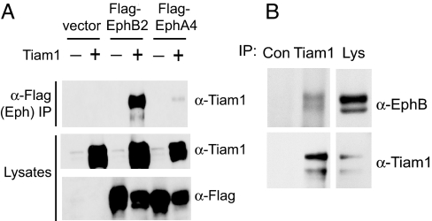
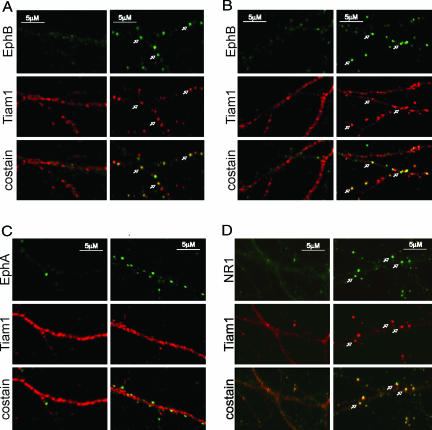
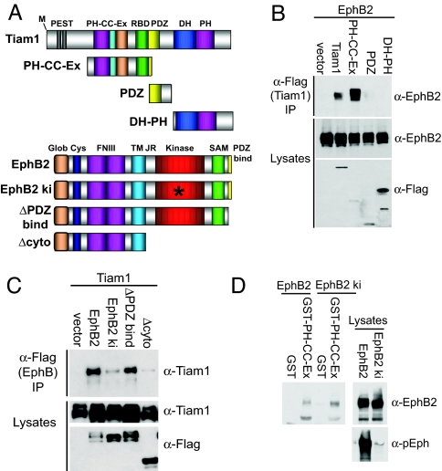
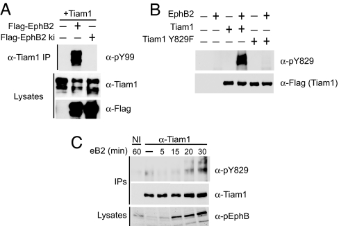
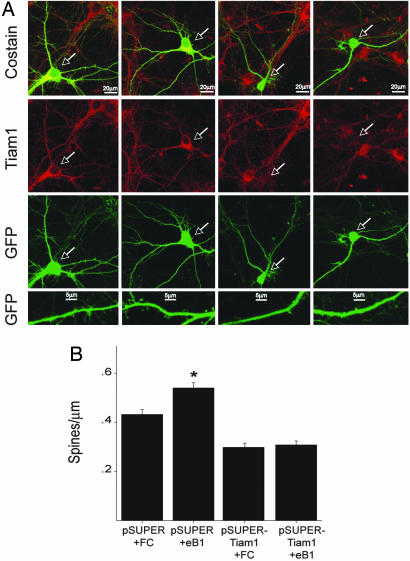
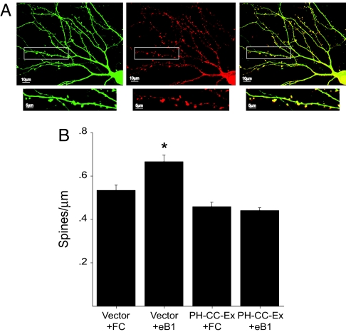
Dendritic spines are small, actin-rich protrusions on the surface of dendrites that receive the majority of excitatory synaptic inputs in the brain. The formation and remodeling of spines, processes that underlie synaptic development and plasticity, are regulated in part by Eph receptor tyrosine kinases. However, the mechanism by which Ephs regulate actin cytoskeletal remodeling necessary for spine development is not fully understood. Here, we report that the Rac1 guanine nucleotide exchange factor Tiam1 interacts with the EphB2 receptor in a kinase-dependent manner. Activation of EphBs by their ephrinB ligands induces the tyrosine phosphorylation and recruitment of Tiam1 to EphB complexes containing NMDA-type glutamate receptors. Either knockdown of Tiam1 protein by RNAi or inhibition of Tiam1 function with a dominant-negative Tiam1 mutant blocks dendritic spine formation induced by ephrinB1 stimulation. Taken together, these findings suggest that EphBs regulate spine development in part by recruiting, phosphorylating, and activating Tiam1. Tiam1 can then promote Rac1-dependent actin cytoskeletal remodeling required for dendritic spine morphogenesis.


Conflict of interest statement

The authors declare no conflict of interest.

Figures

Fig. 1.
Fig. 1.
Tiam1 interacts with EphB2. (A) Extracts of 293T cells transfected with Tiam1 in combination with empty vector, FLAG-EphB2, or FLAG-EphA4 were immunoprecipitated (IP) with anti-FLAG antibodies and then immunoblotted with anti-Tiam1 antibodies. The expression levels of Tiam1, EphB2, and EphA4 were confirmed by immunoblotting (Bottom). (B) Association of endogenous Tiam1 and EphB2. Lysates from P15 rat brain synaptosomes were immunoprecipitated with anti-Tiam1 antibodies or with control (anti-ZAP70) antibodies and then immunoblotted with anti-EphB2 or anti-Tiam1 antibodies. The presence of EphB2 and Tiam1 in the lysate (Lys) was confirmed with anti-EphB2 and anti-Tiam1 antibodies.
Fig. 2.
Fig. 2.
EphrinB stimulation induces the colocalization of Tiam1 with EphB complexes. (A) Tiam1 clusters and colocalizes with EphBs upon ephrinB2 stimulation. E18 rat hippocampal neurons (DIV6) were treated with aggregated ephrinB2-Fc (Right) or control Fc (Left) for 60 min and were then fixed and stained for Tiam1 (red) and EphB (green). The white arrows indicate places where Tiam1 and EphB receptors colocalize. (B) EphrinB1 stimulation induces Tiam1-EphB colocalization. Neurons were stimulated with clustered ephrinB1-Fc (Right) or control Fc (Left) and then fixed and stained for Tiam1 (red) and EphB (green). (C) EphrinA1 stimulation fails to induce Tiam1–EphA colocalization. Neurons were stimulated with aggregated ephrinA1-Fc (Right) or control Fc (Left), fixed, and stained for Tiam1 (red) and EphA (green). (D) Tiam1 colocalizes with NMDA receptors upon ephrinB2 stimulation. E18 rat hippocampal neurons (DIV6) were stimulated with aggregated ephrinB2-Fc (Right) or control Fc (Left), fixed, and stained for Tiam1 (red) and the NR1 subunit of the NMDA receptor (green). The white arrows indicate places where Tiam1 and NR1 colocalize.
Fig. 3.
Fig. 3.
Characterization of the Tiam1–EphB2 interaction. (A) Schematic representation of Tiam1 and EphB2 constructs used in this work. RBD, Ras-binding domain. (B) The Tiam1 PH-CC-Ex domain mediates the interaction with EphB2. EphB2 was cotransfected into 293T cells alone or in combination with one of the following FLAG-tagged Tiam1 constructs: full-length Tiam1, a PH-CC-Ex construct and a Ras-binding domain, a PDZ domain, or the catalytic DH-PH domain. The Tiam1 constructs were immunoprecipitated with anti-FLAG antibodies, and EphB2 coprecipitation was assessed by using anti-EphB2 antibodies. The expression levels of each construct were evaluated by immunoblotting. (C) Tiam1 interacts with the cytoplasmic domain of EphB2 in a kinase-dependent manner. Tiam1 was transiently transfected into 293T cells with one of the following FLAG-tagged EphB2 receptor constructs: full-length EphB2, a kinase-inactive mutant (EphB2 ki), an EphB2 receptor lacking the PDZ-binding domain (ΔPDZ bind), or an EphB2 receptor lacking the entire cytoplasmic domain (Δcyto). The EphB2 receptor constructs were immunoprecipitated (IP) with anti-FLAG antibodies and then immunoblotted with anti-Tiam1 antibodies. The expression levels of each construct were confirmed by immunoblotting. (D) The isolated Tiam1 PH-CC-Ex domain interacts with EphB2 in a kinase-independent manner. Control GST or a GST fusion protein of the Tiam1 PH-CC-Ex domain immobilized on GSH beads was incubated with 293T cell lysate expressing EphB2 or EphB2 ki. Bound proteins were analyzed by immunoblotting with an anti-EphB2 antibody. The levels and activity of the EphB2 receptor constructs in the lysate were assessed with anti-EphB2 and anti-pEphB2 antibodies.
Fig. 4.
Fig. 4.
EphrinB stimulation induces Tiam1 tyrosine phosphorylation. (A) HEK 293T cells were cotransfected with Tiam1 and FLAG-tagged wild-type or kinase-inactive EphB2 receptor, and then Tiam1 was immunoprecipitated (IP) and assessed for tyrosine phosphorylation by using a phosphotyrosine-specific antibody (pY99). Tiam1 and EphB2 receptor expression levels were confirmed with anti-Tiam1 and anti-FLAG antibodies, respectively. (B) Generation of the site-specific phospho-Tiam1 antibody, pY829. FLAG-tagged wild-type and Y829F Tiam1 were expressed in 293T cells alone or in combination with EphB2. The Tiam1 constructs were immunoprecipitated with anti-FLAG antibodies and then examined for phosphorylation on tyrosine-829 by using the phospho-specific Tiam1 antibody, pY829. The failure of the anti-pY829 Tiam1 antibody to recognize Tiam1 Y829F coexpressed with EphB2 suggests that the antibody is site-specific. (C) Tyrosine phosphorylation of Tiam1 in neurons upon ephrinB2-Fc stimulation. E18 hippocampal neurons (DIV4) were treated with clustered ephrinB2-Fc or control Fc for the indicated times, and then Tiam1 was immunoprecipitated with an anti-Tiam1 antibody. Tyrosine phosphorylation of Tiam1 was analyzed by using the anti-pY829 Tiam1 antibody. Immunoprecipitated Tiam1 was visualized with the anti-Tiam1 antibody, and EphB2 receptor autophosphorylation was detected in the lysate by using anti-phospho-EphB2 antibodies.
Fig. 5.
Fig. 5.
RNAi knockdown of Tiam1 expression blocks ephrinB1-induced spine development. (A) Dissociated hippocampal neurons (DIV5) were transfected with an EGFP expression vector together with the pSUPER control vector (columns 1 and 2) or the pSUPER-Tiam1 RNAi vector (columns 3 and 4). Nine days later (DIV14), neurons were stimulated for 4 h with clustered ephrinB1-Fc (eB1) (columns 2 and 4) or control Fc (columns 1 and 3) and then fixed and subjected to immunofluorescence. Tiam1 expression was specifically reduced in neurons transfected with the pSUPER-Tiam1 RNAi: vector (as indicated by the white arrows) but not in neighboring untransfected cells or in neurons transfected with the control pSUPER vector. Suppression of Tiam1 expression inhibited ephrinB1-induced spine development, as can be seen in the bottom row of panels. (B) Quantification of the effect of decreased Tiam1 expression on ephrinB1-induced spine development. ∗, P < 0.001, Student's t test.
Fig. 6.
Fig. 6.
Overexpression of the Tiam1 PH-CC-Ex domain inhibits EphB-dependent spine development. (A) Hippocampal neurons expressing EGFP and myc-tagged PH-CC-Ex domain were fixed and stained for myc. The Tiam1 PH-CC-EX domain (Center) appears to preferentially localize to spines (Right). EGFP (Left). (B) The Tiam1 PH-CC-Ex domain blocks ephrinB1-induced spine development. E18 hippocampal neurons (DIV5) were transfected with an EGFP expression vector together with a myc-tagged PHn-CC-Ex expression vector or an empty control vector. After an additional 9 days in culture (DIV14), neurons were stimulated with aggregated ephrinB1-Fc (eB1) or Fc alone, fixed, and imaged for spine density. ∗, P < 0.001, Student's t test.

References

    1. Harris KM, Kater SB. Annu Rev Neurosci. 1994;17:341–371. - PubMed
    1. Yuste R, Bonhoeffer T. Annu Rev Neurosci. 2001;24:1071–1089. - PubMed
    1. Kaufmann WE, Moser HW. Cereb Cortex. 2000;10:981–991. - PubMed
    1. Kasai H, Matsuzaki M, Noguchi J, Yasumatsu N, Nakahara H. Trends Neurosci. 2003;26:360–368. - PubMed
    1. Yamaguchi Y, Pasquale EB. Curr Opin Neurobiol. 2004;14:288–296. - PubMed

Publication types

MeSH terms