. 2007 Apr 18;2(4):e375.
doi: 10.1371/journal.pone.0000375.

Mechanisms of TSC-mediated control of synapse assembly and axon guidance

Affiliations

Mechanisms of TSC-mediated control of synapse assembly and axon guidance

Sarah Knox et al. PLoS One. .

Abstract

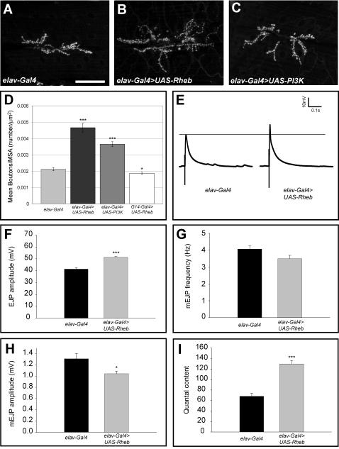
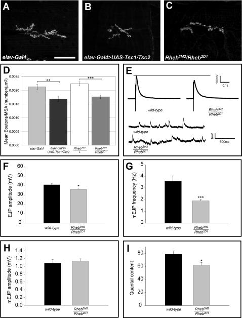
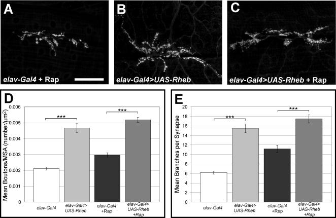
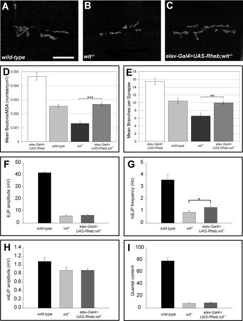
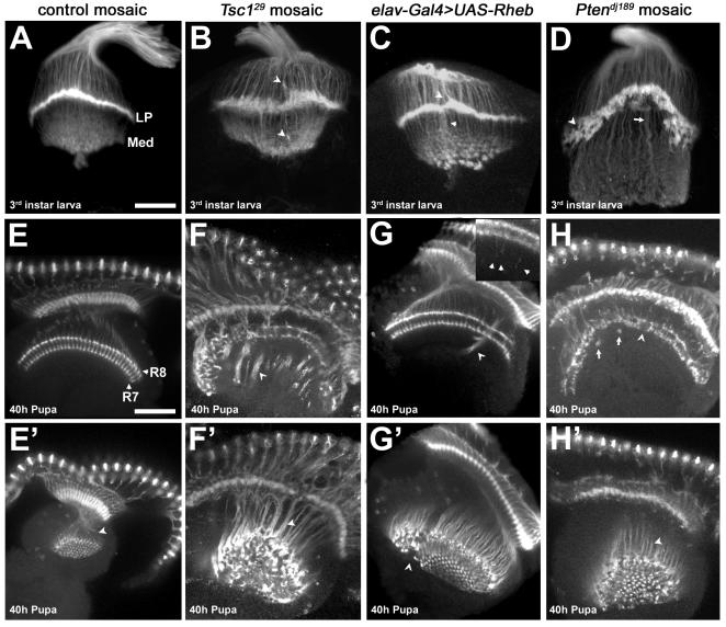
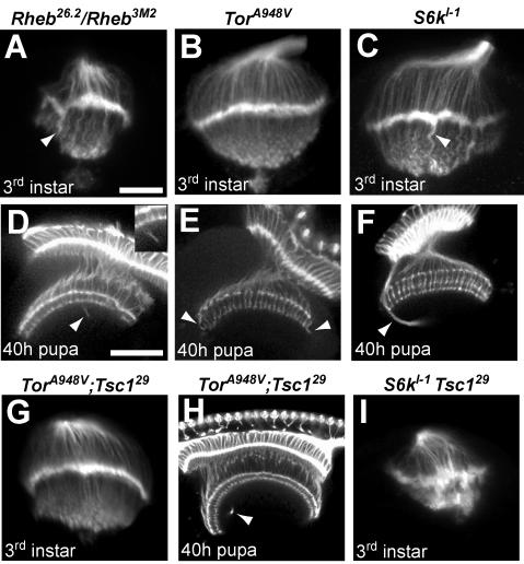
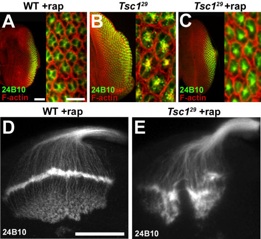
Tuberous sclerosis complex is a dominant genetic disorder produced by mutations in either of two tumor suppressor genes, TSC1 and TSC2; it is characterized by hamartomatous tumors, and is associated with severe neurological and behavioral disturbances. Mutations in TSC1 or TSC2 deregulate a conserved growth control pathway that includes Ras homolog enriched in brain (Rheb) and Target of Rapamycin (TOR). To understand the function of this pathway in neural development, we have examined the contributions of multiple components of this pathway in both neuromuscular junction assembly and photoreceptor axon guidance in Drosophila. Expression of Rheb in the motoneuron, but not the muscle of the larval neuromuscular junction produced synaptic overgrowth and enhanced synaptic function, while reductions in Rheb function compromised synapse development. Synapse growth produced by Rheb is insensitive to rapamycin, an inhibitor of Tor complex 1, and requires wishful thinking, a bone morphogenetic protein receptor critical for functional synapse expansion. In the visual system, loss of Tsc1 in the developing retina disrupted axon guidance independently of cellular growth. Inhibiting Tor complex 1 with rapamycin or eliminating the Tor complex 1 effector, S6 kinase (S6k), did not rescue axon guidance abnormalities of Tsc1 mosaics, while reductions in Tor function suppressed those phenotypes. These findings show that Tsc-mediated control of axon guidance and synapse assembly occurs via growth-independent signaling mechanisms, and suggest that Tor complex 2, a regulator of actin organization, is critical in these aspects of neuronal development.


Conflict of interest statement

Competing Interests: The authors have declared that no competing interests exist.

Figures

Figure 1
Figure 1. Activation of the Tor pathway produces synaptic growth and enhanced physiological function.
The morphology of the third instar larval NMJ was visualized with the presynaptic marker anti-cysteine string protein (CSP) and confocal microscopy. Images shown are stacks of 20 or more optical sections. Neuronal (elav-Gal4) expression of either Rheb (B) or Pi3K (C) increased the size of the synapse compared to control animals bearing the elav-Gal4 transgene alone (A). Numbers of synaptic boutons/muscle area are quantified in D. Expression of either UAS-Rheb (n = 41) or UAS-Pi3K (n = 41) produced a significant increase in the number of boutons/muscle area compared to controls (n = 44), while expression of UAS-Rheb in the muscle (driven by G14-Gal4, n = 18) produced a reduction. Neuron-specific expression of Rheb also produced electrophysiological changes at the NMJ, determined by intracellular recordings from abdominal muscle 6 in third instar larvae. The amplitude of the EJP was significantly increased in animals expressing UAS-Rheb (n = 21) compared to controls with elav-Gal4 alone (n = 12). Examples of EJP voltage traces are shown in E, and mean EJP values are quantified in F. Quantal content, a measure of the number of synaptic quanta released in a single firing of the motoneuron, was nearly doubled by neuron-directed expression of Rheb compared to controls (I). Mini-EJP amplitude was decreased in these animals (H), while mEJP frequency showed no significant change (G). In this and all subsequent figures, *** denotes p-values less than 0.00005 using a student t-test comparison with controls, ** denotes p-values less than 0.005, and * denotes p-values less than 0.05. The scale bar is 50 microns in panel A.
Figure 2
Figure 2. Rheb activity is required for normal synapse assembly.
Panels A–C show anti-CSP staining of larval NMJs in a control animal (A, elav-Gal4 driver alone), an animal bearing elav-Gal4>UAS-Tsc1, UAS-Tsc2 (B), or a Rheb partial loss of function mutant (C). Reduction of Rheb function produced by either neuron-directed expression of Tsc1 and Tsc2 (n = 22) or mutation of Rheb (n = 40) significantly reduced synapse size compared to controls with elav-Gal4 alone (n = 44) or animals heterozygous for a Rheb mutation (n = 17), as measured by the number of synaptic boutons/muscle area (D). Panel E shows sample EJP traces for wild-type and Rheb mutant NMJs, as well as baseline recordings from these preparations showing the size and frequency of mini-EJPs. Panels F, G, and I show reductions in EJP amplitude, mini-EJP frequency, and quantal content for Rheb mutant synapses (n = 29) compared to wild-type controls (n = 10). Mini-EJP amplitude did not show a significant change (H). The scale bar in A is 50 microns.
Figure 3
Figure 3. Rapamycin does not block Rheb-mediated synapse growth.
Panels A–C show anti-CSP staining of NMJ synaptic boutons, demonstrating that the TORC1 inhibitor rapamycin does not block synapse growth in control animals or in larvae with neuron-directed expression of Rheb (elav-Gal4>UAS-Rheb). Panels D and E provide quantification of bouton numbers/muscle area and numbers of motoneuron branches, respectively, for elav-gal4 controls (n = 44), animals with neuronal expression of Rheb (n = 41), control animals treated with rapamycin (n = 26), and Rheb expressing animals treated with rapamycin (n = 29). The scale bar is 50 microns.
Figure 4
Figure 4. Rheb-mediated synapse expansion and physiological function is BMP-signaling dependent.
Anti-CSP staining of synaptic boutons (panels A–C) shows the effects of wit on synapse growth (B), and the effects of neuron-directed expression of Rheb on wit mutant NMJs (C) compared to wild-type (A). Synapse size, measured by either the number of boutons/muscle area (D) or the number of motoneuron branches (E), is dramatically reduced in wit mutants (n = 20) compared to wild-type (n = 12), and is partially rescued by neuron-directed expression of Rheb (elav-Gal4>UAS-Rheb, n = 24). Reductions in EJP amplitudes (F), mini-EJP amplitudes (H), and quantal content (I) mediated by loss of wit (n = 8) are not rescued by neuron-directed expression of Rheb (n = 16) (n = 10 for wild-type). The decrease in mini-EJP frequency of wit mutants, a measure of spontaneous vesicle release, is rescued to a significant degree by expression of Rheb in the motoneuron (G). The scale bar represents 50 microns.
Figure 5
Figure 5. Photoreceptor axon projection defects associated with increased Tor signaling.
(A–D) Dorsal-posterior views of third instar optic lobes stained with MAb24B10 to visualize photoreceptor projections. (A) Mitotic clones in an FRT82B control background show proper termination of photoreceptor axons R1-6 at the lamina plexus (LP), and termination of photoreceptors R7 and R8 in the medulla (Med). (B) Tsc129 mutant axons terminate at incorrect positions above and below the lamina (arrowheads) and produce a broadened lamina plexus. (C) Neuronal expression of Rheb creates axon termination defects similar to those seen in Tsc1 mosaics (D) Ptendj189 mutant photoreceptors leave gaps and holes (arrowhead) in the lamina plexus, which is broader and noticeably “peaked.” The medulla contains axon projections which are thicker and much longer than in controls (arrow). (E–H′) Dorsal view of optic lobes from 40h pupae stained with MAb24B10. E′–H′ are lower optical planes of the optic lobes shown in E–H, respectively. (E, E′) Control photoreceptors R7 and R8 show two distinct layers of termination in the medulla (labels), and are arranged in a highly regular pattern (arrowhead). (F) Animals with Tsc129 mutant photoreceptors show severe disruption of the R7 and R8 termination layers. Instead of terminating at the correct positions, the axons fail to de-fasciculate, forming dense bundles (arrowheads) that project beyond the medulla. (G, G′) Neuron-directed expression of Rheb causes axon bundles to project beyond the medulla in a fashion similar to Tsc1 mosaics (arrowheads), but the phenotype is much less severe. (G, inset) Individual Rheb-overexpressing axons show an intermediate termination defect, stopping several microns beyond their normal targets (arrowheads in inset). (H) Ptendj189 mutant axons exhibit gaps and collapses in the R7/R8 termination zone (arrowhead). Thick axon bundles can be seen that bypass their usual stopping points and then loop back to terminate at other locations in the R7/R8 layers (arrows). (H′, F′) Axon bundles in Ptendj189 mosaics are not as densely packed as those of Tsc129 mosaics (arrowheads), but are still disorganized. All scale bars are 50 microns.
Figure 6
Figure 6. Effects of mutations that downregulate the Tor pathway on photoreceptor axon guidance, and genetic epistasis with Tsc1.
Optic lobes from third instar larvae (A–C) and 40h pupae (D–F) stained with MAb24B10. (A) Larvae heteroallelic for a hypomorphic combination of Rheb alleles show abnormal photoreceptor patterning and contain thick axon bundles that extend into the medulla (arrowhead). (D) At the 40 h pupal stage, Rheb mutants display axons that bypass their normal targets in the R7/R8 termination zones (arrowhead). (B) Larvae homozygous for a hypomorphic Tor allele show fairly normal photoreceptor patterning, but at the pupal stage (E) misrouted axons can be seen in the medulla (arrowheads). (C) S6k null homozygous larvae show thick axon bundles projecting past the lamina (arrowhead), while S6k pupae (F) display misrouted axons that initially bypass the R7/R8 termination zone (arrowhead). (G, H) Animals doubly mutant for Tor and Tsc1 do not show the severe photoreceptor defects seen when axons are mutant for Tsc1 alone (compare to Figure 5B, F, F′), although mild defects similar to those in Tor mutants are still apparent (arrowhead). (I) S6k-Tsc1 double homozygous mutants display a severe phenotype dissimilar to mutants for either S6k or Tsc1 alone. The scale bar is 25 microns in panel A, 50 microns in panel D.
Figure 7
Figure 7. Axon guidance defects in Tsc1 mosaics are not suppressed by blocking growth.
(A–C) Third instar eye discs from wild type and Tsc1 mosaic larvae raised with or without rapamycin (rap). Ommatidial units, comprised of eight photoreceptors, were visualized with phalloidin (red) that detects F-actin, and MAb24B10 (green). Phalloidin staining is strongest at the perimeter of each ommatidium, outlining each sensory unit. Rapamycin treatment of Tsc1 mosaic eye discs (C) restored eye disk size and cell size compared to wild type (A). (D and E) Rapamycin treated third instar larval brains stained with MAb24B10. Rapamycin treatment blocked abnormal growth of the retina and the increase in photoreceptor cell size, but did not ameliorate the abnormal axon projections also characteristic to untreated Tsc129 mosaics. The scale bars in panel A represent 50 microns in the left image, 10 microns in the right image. The scale bar is 50 microns in panel D.

References

    1. Ess KC. The neurobiology of tuberous sclerosis complex. Semin Pediatr Neurol. 2006;13:37–42. - PubMed
    1. Tavazoie SF, Alvarez VA, Ridenour DA, Kwiatkowski DJ, Sabatini BL. Regulation of neuronal morphology and function by the tumor suppressors Tsc1 and Tsc2. Nat Neurosci. 2005;8:1727–1734. - PubMed
    1. von der Brelie C, Waltereit R, Zhang L, Beck H, Kirschstein T. Impaired synaptic plasticity in a rat model of tuberous sclerosis. Eur J Neurosci. 2006;23:686–692. - PubMed
    1. Acebes A, Ferrus A. Increasing the number of synapses modifies olfactory perception in Drosophila. J Neurosci. 2001;21:6264–6273. - PMC - PubMed
    1. Canal I, Acebes A, Ferrus A. Single neuron mosaics of the drosophila gigas mutant project beyond normal targets and modify behavior. J Neurosci. 1998;18:999–1008. - PMC - PubMed

Publication types