. 2007 Jun 1;306(1):208-21.
doi: 10.1016/j.ydbio.2007.03.018. Epub 2007 Mar 20.

The Hectd1 ubiquitin ligase is required for development of the head mesenchyme and neural tube closure

Affiliations

The Hectd1 ubiquitin ligase is required for development of the head mesenchyme and neural tube closure

Irene E Zohn et al. Dev Biol. .

Abstract

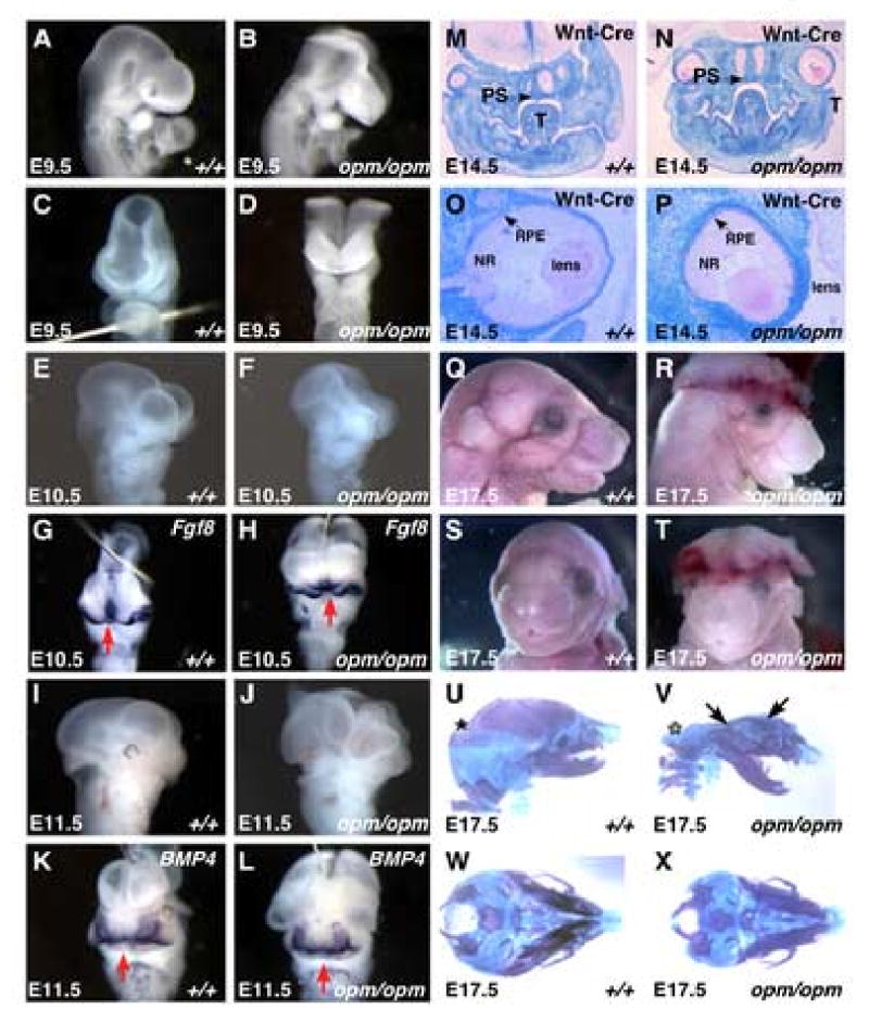
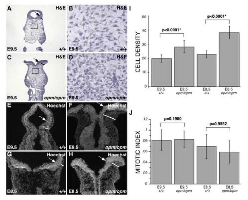
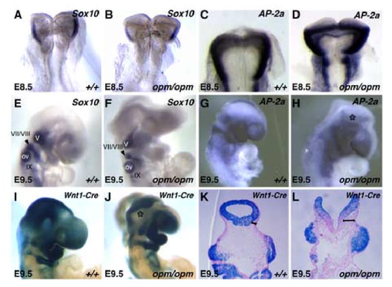
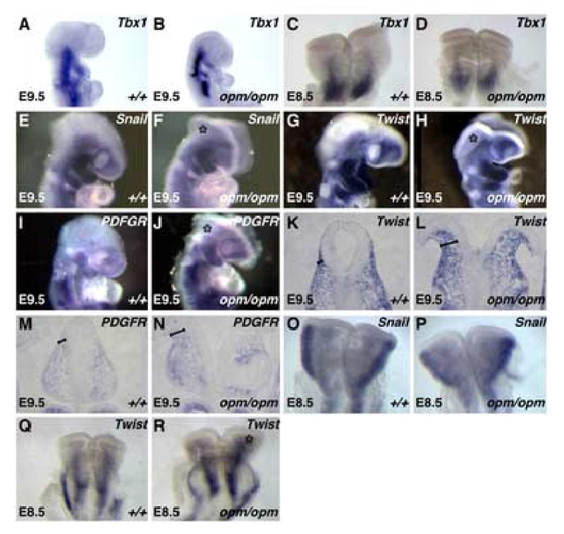
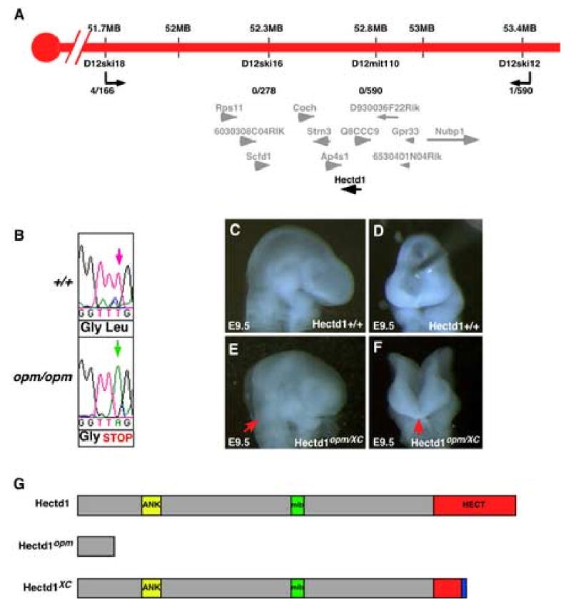
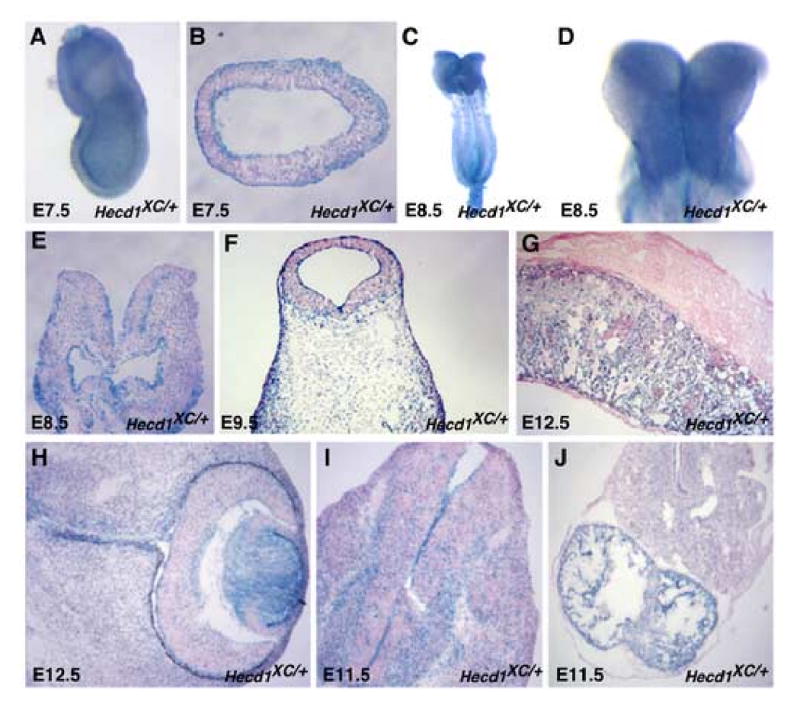
Closure of the cranial neural tube depends on normal development of the head mesenchyme. Homozygous-mutant embryos for the ENU-induced open mind (opm) mutation exhibit exencephaly associated with defects in head mesenchyme development and dorsal-lateral hinge point formation. The head mesenchyme in opm mutant embryos is denser than in wildtype embryos and displays an abnormal cellular organization. Since cells that originate from both the cephalic paraxial mesoderm and the neural crest populate the head mesenchyme, we explored the origin of the abnormal head mesenchyme. opm mutant embryos show apparently normal development of neural crest-derived structures. Furthermore, the abnormal head mesenchyme in opm mutant embryos is not derived from the neural crest, but instead expresses molecular markers of cephalic mesoderm. We also report the identification of the opm mutation in the ubiquitously expressed Hectd1 E3 ubiquitin ligase. Two different Hectd1 alleles cause incompletely penetrant neural tube defects in heterozygous animals, indicating that Hectd1 function is required at a critical threshold for neural tube closure. This low penetrance of neural tube defects in embryos heterozygous for Hectd1 mutations suggests that Hectd1 should be considered as candidate susceptibility gene in human neural tube defects.


Figures

Figure 1
Figure 1. opm mutant embryos exhibit defects in neural tube closure without other significant defects in craniofacial development
Panels A-L: lateral (A, B, E, F, I and J) and frontal (C, D, G, H, K and L) views of wildtype (A, C, E, G, I and K) and opm mutant (B, D, F, H, J, L) heads demonstrating neural tube closure defect from the forebrain to the hindbrain and normal development of the face (A-D, E9.5; E-H, E10.5; I-L, E11.5). Frontal views of wildtype and opm mutant embryos at E10.5 and E11.5 were labeled by in situ hybridization using probes against Fgf8 and BMP4 respectively to highlight the developing frontal nasal processes. Fate of the neural crest as shown in coronal sections of E14.5 Wnt1-Cre/R26R wildtype (M,O) and opm mutant (N,P) embryos stained for β-galactosidase activity and counterstained with eosin. Arrow points to the properly fused secondary palatal shelves (PS; T=Tongue) in wildtype (M) and opm mutant (N) heads. Eye development is abnormal in opm mutant embryos (P). The retinal pigmented epithelium (RPE, arrow), neural retina (NR) and lens appear histologically normal, but eyes are rotated in the head of opm mutant embryos. Profile (Q,R) and frontal (S,T) views of E17.5 wildtype (Q,S) and opm mutant (R,T) heads demonstrating normal craniofacial development in spite of the severe neural tube closure defect. Lateral (U,V) and basal (W,X) views of E17.5 skeletal preparations stained with Alcian blue and Alizarin Red in wildtype (U,W) and opm mutant (V,X) skulls. Asterisk (*) denotes exoccipital, petrosal and interpariatal bones that are missing in opm mutant skulls. Arrows point to frontal and pariatal bones that are malformed in opm mutant skulls.
Figure 2
Figure 2. Abnormal head mesenchyme development in opm mutant embryos is associated with neural tube closure defects
A-D. Hematoxylin and Eosin (H&E) staining of coronal sections of wildtype (A,B) and opm mutant (C,D) heads at E9.5. Brackets highlight the region of head mesenchyme that are expanded around the opm neural tube. Panels B&D show magnified views of the boxes drawn in panels A&C, demonstrating the abnormally dense cellular organization of opm mutant head mesenchyme. E-H. Nuclei were visualized by staining with Hoechst in coronal sections of wildtype (E,G) and opm mutant (F,H) at E9.5 (E,F) and E8.5 (G,H) demonstrating that the head mesenchyme is denser around the dorsal neural tube of opm mutant embryos during neurulation. There is also a failure of dorsal-lateral hinge point formation leading to a flat or convex curvature of the opm neural tube compared to the concave neural tube in wildtype embryos at both stages. Arrows point to dorsal-lateral hinge points in A-H. I. Cell density was determined by counting the number of nuclei in 1-2 defined areas on many sections from many embryos (E9.5: wildtype n=39 areas, opm mutant n=41 areas; E8.5: wildtype n=58 areas, opm mutant n=63 areas). Error bars represent one standard deviation from the mean. The density of cells between wildtype and opm mutant samples are significantly different as calculated by the Mann-Whitney U test (E9.5: P-value<0.0001*; E8.5: P-value<0.0001*). J. Mitotic indexes were calculated by dividing the number of P-H3 positive cells by the number of nuclei in a given area for wildtype and opm mutant in two to three sections from many embryos (E9.5: wildtype n=19 sections, opm mutant n=20 sections; E8.5: wildtype n=17 sections, opm mutant n=18 sections). Error bars represent one standard deviation from the mean. Samples are not significantly different as calculated by the Mann-Whitney U test (E9.5: P-value=0.9552; E8.5: P-value=0.1980).
Figure 3
Figure 3. Neural crest cells are not present in the abnormal opm mutant head mesenchyme
E8.5 (A-D) and E9.5 (F-H) wildtype (A,C,E,G) and opm mutant (B,D,F,H) embryos were analyzed by whole-mount in situ hybridization analysis for expression of Sox10 (A,B,E,F) and AP-2α (C,D,G,H). Asterisk (*) in panel H denotes the lack of AP-2α expression in the abnormal head mesenchyme surrounding the open neural tube in the opm mutant head. V, VII/VIII and IX denote the Sox10-expressing cranial nerves. OV = otic vesicle. Fate of the neural crest in E9.5 Wnt1-Cre/R26R wildtype (I) and opm mutant (J) embryos that were stained in whole-mount for β-galactosidase activity. Asterisk (*) highlights the abnormal head mesenchyme in opm mutant embryos that is negative for β-galactosidase activity. Coronal sections of E9.5 Wnt1-Cre/R26R wildtype (K) and opm mutant (L) head stained for β-galactosidase and counterstained with eosin. Brackets highlight the abnormally expanded head mesenchyme in opm mutant embryos that is negative for β-galactosidase activity.
Figure 4
Figure 4. The abnormal head mesenchyme in opm mutant embryos expresses molecular markers of cephalic mesoderm
E9.5 (A,B,E,F,G,H,I,J) and E8.5 (C,D,O,P,Q,R) wildtype (A,C,E,G,I,O,Q) and opm mutant (B,D,F,H,J,P,R) heads were analyzed by whole-mount in situ hybridization analysis for expression of Tbx1 (A-D), Snail (E,F,O,P), Twist (G,H,Q,R) and PDGFRα (I,J). Asterisk (*) denotes staining in the abnormal head mesenchyme surrounding the open neural tube in opm mutant embryos. Coronal sections of wildtype (K,M) and opm mutant (L,N) heads stained by in situ hybridization for expression of Twist (K,L) and PDGFRα (M,N). Brackets highlight the abnormal head mesenchyme surrounding the open neural tube in opm mutant embryos.
Figure 5
Figure 5. The opm mutation disrupts the uncharacterized Hectd1 ubiquitin ligase
A. Genetic map of opm interval on mouse chromosome 12. The number of recombination events over the number of opportunities for recombination is indicated for each polymorphic marker. Markers D12ski16 and D12MIT110 never separated from the opm phenotype. Within this interval are twelve transcription units: Rps11 (ribosomal protein S11), 6030308C04RIK (RIKEN cDNA 6030408C04 gene), Scfd1 (sec1 family domain containing 1), Coch (coagulation factor C homolog), Strn3 (striatin, calmodulin binding protein 3), Ap4s1 (adaptor-related protein complex AP-4, sigma 1), Hectd1 (HECT domain containing 1), Q8CCC9 (PREDICTED: hypothetical protein), D930036F22Rik (RIKEN cDNA D930036F22 gene), 6530401N04Rik (RIKEN cDNA 6530401N04 gene), Gpr33 (G protein-coupled receptor 33) and Nubp1 (nucleotide binding protein-like). The transcripts in the opm interval and physical map position (mb) is from the Ensemble mouse genome assembly release #40. B. The opm ENU-induced mutation results in a T to A transversion (green arrow) at position 430 in the Hectd1 coding sequence. This mutation results in a nonsense mutation changing a Leucine to a stop codon. C-F. The Hectd1XC gene trap allele fails to complement Hectd1opm as embryos at E9.5 exhibit exencephaly from the hindbrain to the forebrain (E,F). Panels C-F show lateral (C,E) and frontal (D,F) views of wildtype (C,D) and Hectd1opm/XC mutant (E,F) E9.5 embryos. G. Predicted protein motifs in Hectd1: an ankyrin domain (ANK), MIB-HERC2 domain (mib) and a C-terminal Homologous to the E6-AP Carboxyl Terminus domain (HECT). The Hectd1opm mutation results in a truncated protein at amino acid 145. The Hectd1XC mutation results in an insertion and truncation of the HECT domain.
Figure 6
Figure 6. Hectd1 is ubiquitously expressed during development of the mouse embryo
Hectd1 expression was monitored by staining for β-galactosidase activity in whole mount (A,C,D) or in section and counterstained with eosin (B,E-J) in Hectd1XC/+ embryos. Hectd1 is expressed during development of the head mesenchyme at E7.5 (A,B), E8.5 (C-E) E9.5 (F). Hectd1 is also expressed in the placenta (G) and eye (H) at E12.5. Hectd1 is expressed at E11.5 at higher levels in differentiated neurons of the developing spinal cord (I) and the atrium of the heart (J).

References

    1. Bernard DJ, Burns KH, Haupt B, Matzuk MM, Woodruff TK. Normal reproductive function in InhBP/p120-deficient mice. Mol Cell Biol. 2003;23:4882–91. - PMC - PubMed
    1. Brewer S, Feng W, Huang J, Sullivan S, Williams T. Wnt1-Cre-mediated deletion of AP-2alpha causes multiple neural crest-related defects. Dev Biol. 2004;267:135–52. - PubMed
    1. Caspary T, Anderson KV. Patterning cell types in the dorsal spinal cord: what the mouse mutants say. Nat Rev Neurosci. 2003;4:289–97. - PubMed
    1. Chapman DL, Garvey N, Hancock S, Alexiou M, Agulnik SI, Gibson-Brown JJ, Cebra-Thomas J, Bollag RJ, Silver LM, Papaioannou VE. Expression of the T-box family genes, Tbx1-Tbx5, during early mouse development. Dev Dyn. 1996;206:379–90. - PubMed
    1. Chapman SC, Bernard DJ, Jelen J, Woodruff TK. Properties of inhibin binding to betaglycan, InhBP/p120 and the activin type II receptors. Mol Cell Endocrinol. 2002;196:79–93. - PubMed

Publication types

MeSH terms