. 2007 Jun 15;395(1-2):125-34.
doi: 10.1016/j.gene.2007.02.026. Epub 2007 Mar 16.

Regional FMRP deficits and large repeat expansions into the full mutation range in a new Fragile X premutation mouse model

Affiliations

Regional FMRP deficits and large repeat expansions into the full mutation range in a new Fragile X premutation mouse model

Ali Entezam et al. Gene. .

Abstract

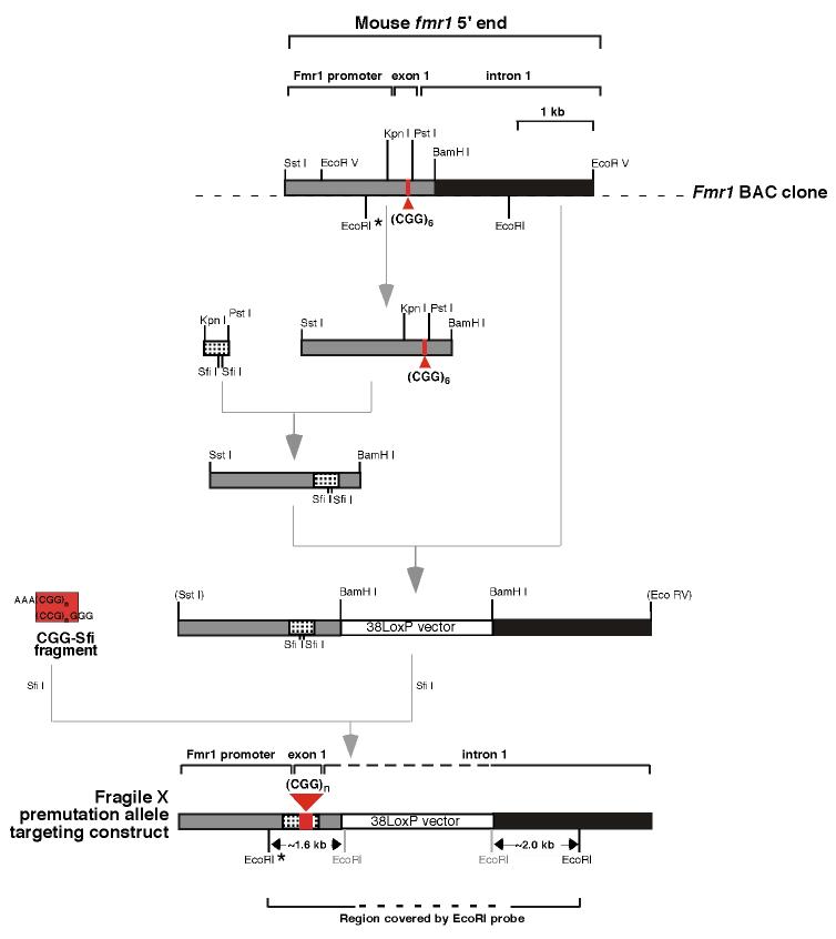
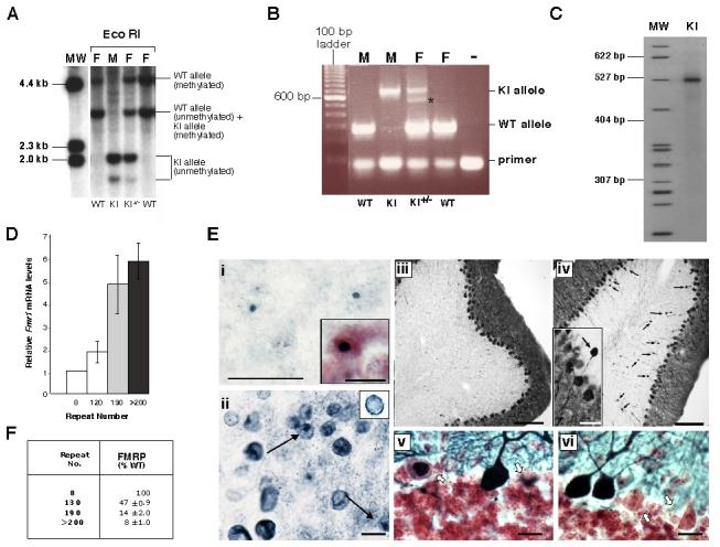
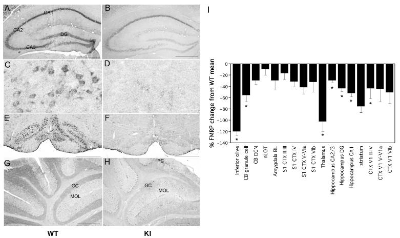
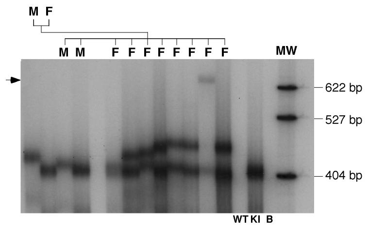
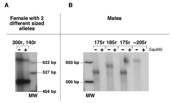
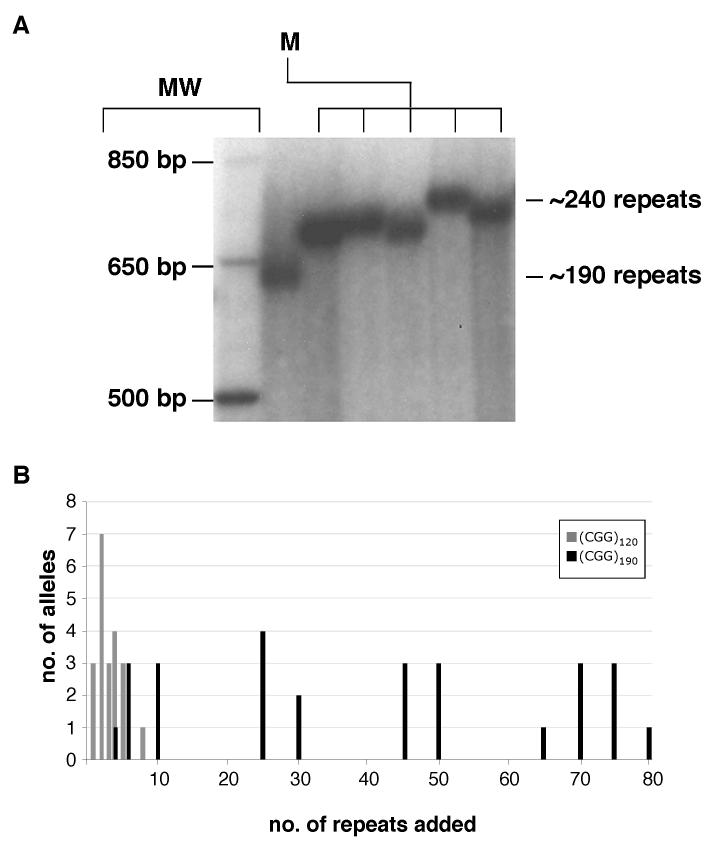
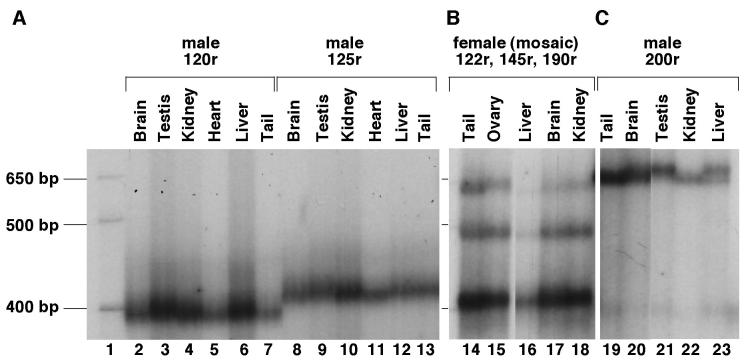
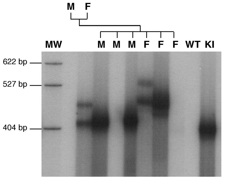
Carriers of FMR1 alleles with 55-200 repeats in the 5' UTR are at risk for Fragile X associated tremor and ataxia syndrome. The cause of the neuropathology is unknown but is thought to be RNA-mediated. Maternally transmitted premutation alleles are also at risk of expansion of the repeat tract into the "full mutation" range (>200 repeats). The mechanism responsible for expansion is unknown. Full mutation alleles produce reduced amounts of the FMR1 gene product, FMRP, which leads to Fragile X mental retardation syndrome. We have developed a murine model for Fragile X premutation carriers that recapitulates key features seen in humans including a direct relationship between repeat number and Fmr1 mRNA levels, an inverse relationship with FMRP levels and Purkinje cell dropout that have not been seen in a previously described knock-in mouse model. In addition, these mice also show a differential deficit of FMRP in different parts of the brain that might account for symptoms of the full mutation that are seen in premutation carriers. As in humans, repeat instability is high with expansions predominating and, for the first time in a mouse model, large expansions into the full mutation range are seen that occur within a single generation. Thus, contrary to what was previously thought, mice may be good models not only for the symptoms seen in human carriers of FMR1 premutation alleles but also for understanding the mechanism responsible for repeat expansion, a phenomenon that is responsible for a number of neurological and neurodevelopmental disorders.


Figures

Fig. 1
Fig. 1. Outline of targeting vector construction strategy
The Kpn I-Pst I fragment that includes exon 1 of the WT murine Fmr1 gene was modified so as to replace the normal mouse CGG•CCG-repeats with 2 different Sfi I sites. This modified fragment was substituted for the WT Kpn I-Pst I fragment in a Bam HI-Sst I subclone of the 5' end of the Fmr1 gene. After the insertion of the resultant modified 5' arm (grey rectangle) and the 3' arm (black rectangle) into the 38LoxP cloning vector (white rectangle), the construct was digested with Sfi I and ligated to the CGG•CCG-repeat fragment that was generated as previously described (Lavedan et. al., 1998) and was flanked by Sfi I sites compatible with those introduced into exon 1. Restriction enzyme sites that are abolished on cloning are shown in parentheses. Sites introduced into the targeting construct from vector derived E. coli sequences are shown in grey font.
Fig. 2
Fig. 2. Characterization of premutation mice generated with the targeting construct shown in Fig. 1
A) Southern blot analysis of Eco RI digested genomic DNA from the tails of WT and KI animals using a probe containing the Eco RI fragment that spans the CGG•CCG-repeat in WT mice (shown at the bottom of Fig. 1). MW, molecular weight marker; M, male; F, female. B) PCR genotyping of mice using frax-c and frax-f primers. Note that in reactions with DNA from females heterozygous for the KI allele, 2 bands are seen in the region expected for the KI allele. Only one band is seen on denaturing gels. The smaller of the 2 bands (indicated by the asterisk) results from a heteroduplex formed between the KI and WT alleles in the last round of the PCR. C) Determination of the repeat size in KI mice using primers frax-m4 and frax-m5. MW, molecular weight marker. D) Relative Fmr1 mRNA levels in 2 month old WT and KI male mice. The difference in RNA levels between WT and KI animals were significant at p<0.0001 by Students t-test. E) CNS pathology in male premutation mice with 120 repeats at ∼100 weeks of age. i) Ubiquitin immunoreactivity in the parafascicular nucleus of the thalamus. (Inset: a section from the reticular formation counterstained with Neutral Red showing a larger inclusion). ii) Lamin immunoreactivity in the nodulus of the cerebellum showing lamin-positive inclusions (arrows) and the irregular distribution of lamin (Inset: cell from WT mice showing the normal lamin distribution on the nuclear periphery) iii-vi) Calbindin immunoreactivity in the cerebellum. iii-iv) Calbindin staining in the cerebellum of WT (iii) and KI (iv) mice. Note that the KI mouse has increased numbers of torpedo axon swellings (arrows in iv). Some are almost as large as the Purkinje soma (iv, inset). v-vi) Cerebellum from KI mice stained for calbindin in blue/black and counterstained with Neutral Red showing Purkinje cells with abnormal calbindin distribution (arrows). In panels i, ii , v and vi the bars indicate 10 μm. The bars in panel iii and iv indicate 100 μm, and the bar in iv (inset) indicates 25 μm. F) Relative FMRP levels in 2 month old WT and KI male mice based on Western blots with FMRP 7G1-1 antibody and normalized using Actin antibodies as described in the text. The data presented represent the results of 3 independent experiments. The difference in protein levels in WT and KI animals were significant at p<0.0001 by Students t-test.
Fig 3
Fig 3. Regional FMRP deficits in male premutation mice with 120 repeats
A-H). Micrographs showing the FMRP localization in the hippocampal complex (A and B), the ventral posteriolateral thalamus (C and D), the inferior olive (E and F) and the cerebellum (G and H) from 2 year old WT (A, C, E, and G) and KI male mice with 120 repeats (B, D, F, and H) mice. Bars = 500 μm for A, B, E, F, G and H and 25 μm for C and D. GC, granule cells; MOL: molecular layer, PC: Purkinje cells. I). Bar graph showing the degree of change in FMRP expression in KI relative to WT. BL, basolateral; CB, cerebellum; DCN, deep cerebellar nuclei; CTX, cortex; DG, dentate gyrus; nLOT, nucleus of lateral olfactory tract. * Significantly different from WT p<0.05. The data represent the average obtained from 4 WT males and 5 KI males all ∼100 weeks in age.
Fig. 4
Fig. 4. Detection of a large expansion that generates an allele in the full mutation range on germline transmission
The repeat size in a breeding pair involving a male with a premutation allele of ∼120 repeats and a female homozygous for the same sized repeat and one of their litters showing one expansion into the full mutation range indicated by the arrow. MW, molecular weight marker, M, male, F, female, KI, PCR positive control (DNA from a mouse previously shown to carry the premutation), B, PCR negative control (DNA from a WT animal),
Fig. 5
Fig. 5. Analysis of the methylation status of mice with different numbers of repeats
Tail DNA from mice taken at weaning was digested with Sau96 I and then amplified by PCR using primers frax-m4 and frax-m5 that overlap the Sau96 I sites. The PCR products were then compared to the PCR products generated from equivalent amounts of tail DNA that had not been digested with Sau96 I. MW, molecular weight marker
Fig. 6
Fig. 6. Paternal transmission of an allele with 190 repeats
A) PCR analysis of a male mouse with 190 repeats (M) and the progeny produced when this mouse was crossed to a WT female. MW, molecular weight marker, B) Comparison of allele sizes in the progeny of male mice with 120 and 190 repeats. The gray bars represent the number of alleles of the indicated size seen in the progeny of a cross between a male with 120 repeats and a WT female. The black bars indicate the same data obtained from a cross between a male with 190 repeats and a WT female.
Fig. 7
Fig. 7. Somatic stability of premutation alleles
PCR products containing the repeat were generated from tail DNA of mice taken 3 weeks after birth, and the DNA from the indicated organs of adult animals 6-18 months old. The sex of the mice, the number of repeats in their tail DNA and the organ from which the DNA was derived, are indicated above the gel. The samples shown in lanes 14-18 are from a female who inherited 2 premutation alleles and is mosaic for one of them.
Fig. 8
Fig. 8. Germline transmission of alleles from a female mosaic for the premutation
PCR analysis of the repeat size in a mosaic but heterozygous female, the WT male to which she was bred and 1 litter from this cross. MW, molecular weight marker, M, male, F, female, -, PCR negative control (DNA from a WT animal), +, PCR positive control (DNA from a mouse previously shown to carry the premutation). Note that one of the offspring of this cross, the female shown in lane 7, is also mosaic. The mice in lanes 5 and 9 inherited the normal maternal allele as assessed by PCR using primers that amplify both WT and KI alleles (data not shown).

References

    1. Aziz M, Stathopulu E, Callias M, Taylor C, Turk J, Oostra B, Willemsen R, Patton M. Clinical features of boys with fragile X premutations and intermediate alleles. Am J Med Genet B Neuropsychiatr Genet. 2003;121:119–27. - PubMed
    1. Bontekoe CJ, Bakker CE, Nieuwenhuizen IM, van der Linde H, Lans H, de Lange D, Hirst MC, Oostra BA. Instability of a (CGG)98 repeat in the Fmr1 promoter. Hum Mol Genet. 2001;10:1693–9. - PubMed
    1. Brouwer JR, Mientjes EJ, Bakker CE, Nieuwenhuizen IM, Severijnen LA, Van der Linde H, Nelson DL, Oostra BA, Willemsen R. Elevated Fmr1 mRNA levels and reduced protein expression in a mouse model with an unmethylated Fragile X full mutation. Experimental Cell Research. 2006 In press. - PMC - PubMed
    1. Brown V, Jin P, Ceman S, Darnell JC, O'Donnell WT, Tenenbaum SA, Jin X, Feng Y, Wilkinson KD, Keene JD, Darnell RB, Warren ST. Microarray identification of FMRP-associated brain mRNAs and altered mRNA translational profiles in fragile X syndrome. Cell. 2001;107:477–87. - PubMed
    1. Dutch-Belgian Fragile X Consortium Fmr1 knockout mice: a model to study fragile X mental retardation. Cell. 1994;78:23–33. - PubMed

Publication types

MeSH terms