. 2007 Jun;26(3):558-68.
doi: 10.1016/j.nbd.2007.02.007. Epub 2007 Feb 23.

Is functional state of spinal microglia involved in the anti-allodynic and anti-hyperalgesic effects of electroacupuncture in rat model of monoarthritis?

Affiliations

Is functional state of spinal microglia involved in the anti-allodynic and anti-hyperalgesic effects of electroacupuncture in rat model of monoarthritis?

Sun Shan et al. Neurobiol Dis. 2007 Jun.

Abstract

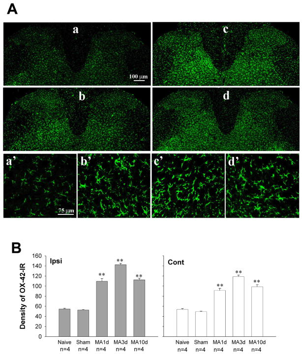
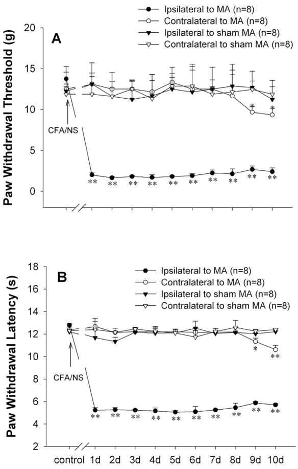
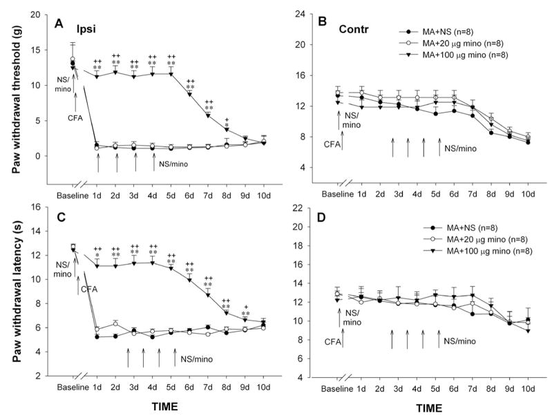
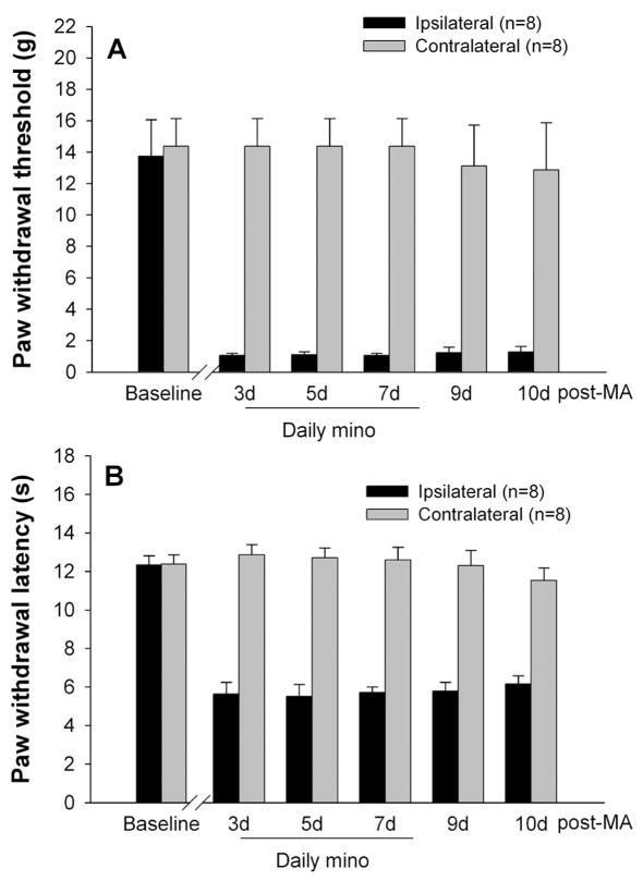
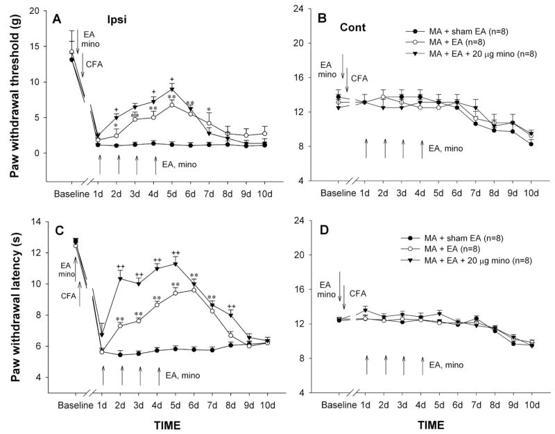
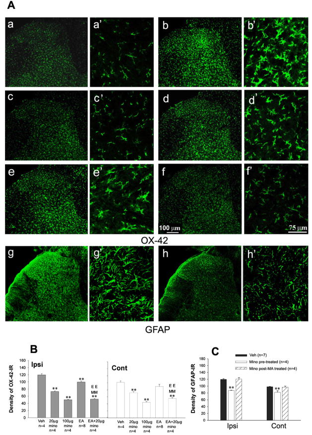
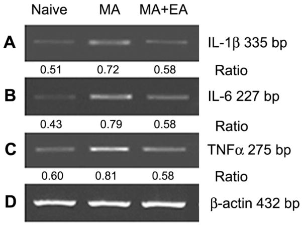
Spinal microglia play a key role for creating exaggerated pain following tissues inflammation or injury. Electroacupuncture (EA) can effectively control the exaggerated pain both in humans with inflammatory disease and animals with experimental inflammatory pain. However, little is known about the relationship between spinal glial activation and EA analgesia. Using immunohistochemistry, RT-PCR analysis, and behavioral testing, the present study demonstrated that (1) Unilateral intra-articular injection of CFA produced a robust microglial activation and the up-regulation of the tumor necrosis factor (TNF)-alpha, interleukin (IL-1beta), and IL-6 mRNA levels in the spinal cord; (2) Repeated intrathecal (i.t.) injection of minocycline (100 microg), a microglial inhibitor, or EA stimulation of ipsilateral "Huantiao"(GB30) and "Yanglingquan" (GB34) acupoints significantly suppressed CFA-induced nociceptive behavioral hypersensitivity and spinal microglial activation; (3) Combination of EA with minocycline significantly enhanced the inhibitory effects of EA on allodynia and hyperalgesia. For the first time, these data provide direct evidence for the involvement of spinal microglial functional state in anti-nociception of EA. Thus, anti-neuroinflammatory effect of EA might be considered as one of the mechanisms of its anti-arthritic pain effects, and thereby a multidisciplinary integrated approach to treating symptoms related to arthritis might be raised.


Figures

Fig. 1
Fig. 1
Time course of OX-42 (microglial marker) expression on the lumbar spinal cord following monoarthritis (MA). (A) Photomicrographs showing OX-42-immunoreactivity (OX-42-IR) on both sides of the spinal dorsal horn on day 3 post-sham MA (a, a′), and day 1(b, b′), day 3 (c, c′), day 10 (d, d′) post-MA. (Aa–d) 10 ×; (Aa′–d′) 40 ×. (B) Quantification of OX-42-IR showing significant up-regulation in all time-point examined. ** p<0.01 vs. sham MA.
Fig. 2
Fig. 2
Unilateral intra-articular injection of complete Freund’s adjuvant (CFA) induced significant mechanical allodynia (A) and thermal hyperalgesia (B) initially in the ipsilateral and subsequently in the contralateral hindpaw. **p<0.01 vs. before CFA injection.
Fig. 3
Fig. 3
Effects of repeated intrathecal (i.t.) injections of minocycline (mino) at different doses on PWT (A & B) and PWL (C & D) of hindpaws ipsilateral (Ipsi, A & C) and contralateral (Cont, B & D) to MA. Repeated i.t. normal saline (NS) or minocycline were given once daily for 5 days with the first injection 30 min before MA (preemptive treatment), and behavioral testes were performed immediately before i.t. injection every day. * p<0.05, ** p<0.01 vs. NS; + p<0.05, ++ p<0.01 vs. 20 μg minocycline.
Fig. 4
Fig. 4
Effects of repeated intrathecal (i.t.) injections of 100 μg minocycline (mino) on PWT (A) and PWL (B) of both hindpaws. Repeated i.t. minocycline were administered once daily for 5 days starting on day 3 after monoarthritis (MA) (post-MA treatment), and behavioral tests were performed immediately before i.t. injections.
Fig. 5
Fig. 5
Effects of repeated sham electroacupuncture (EA), EA, or EA plus i.t. minocycline (mino, 20 μg) on PWT (A & B) and PWL (C & D) of the hindpaws ipsilateral (Ipsi, A & C) and contralateral (Cont, B & D) to monoarthritis (MA). Sham EA, EA, and EA plus i.t. minocycline were given once daily for 5 days with the first injection 30 min before MA, and behavioral tests were performed immediately before EA or EA plus i.t. minocycline every day. * p<0.05, ** p<0.01 vs. sham EA; + p<0.05, ++ p<0.01 vs. EA alone.
Fig. 6
Fig. 6
Effects of repeated intrathecal (i.t.) injections of minocycline (mino), electroacupuncture (EA), co-application of EA and 20 μg minocycline on increase in OX-42-IR (and GFAP-IR) expression on the spinal dorsal horn induced by monoarthritis (MA). (A) Photomicrographs showing OX-42-IR (Aa–f) and GFAP-IR (Ag & Ah) on the ipsilateral spinal dorsal horn by 10 days post-MA. (Aa, a′) Naïve; (Ab,b′ & Ag, g′) Vehicle (Veh); (Ac,c′ & h,h′) 100 μg minocycline; (Ad, d′) 20 μg minocycline; (Ae,e′) EA; (Af, f′) 20 μg minocycline plus EA. (Aa–h) 10 ×; (Aa′–h′) 40 ×. (B) Quantification of OX-42-IR showing that repeated i.t. minocycline and EA suppressed OX-42-IR expression on the spinal dorsal horn. Repeated EA, i.t. minocycline and minocycline plus EA were given once daily for 5 days with the first administration 30 min before MA on day 0 (days 0–4). (C) Quantification of GFAP-IR showing that repeated i.t. minocycline (pre-treatment) suppressed GFAP-IR expression on the spinal dorsal horn. Rats were perfused for immunohistochemistry analysis on day 10 after behavioral tests. * p<0.05, ** p<0.01 vs. vehicle control; EE p<0.01 vs. EA alone; MM p<0.01 vs. 20 μg minocycline alone. mino pre-treated, repeated i.t. minocycline was given once daily for 5 days with the first administration 30 min before MA on day 0; mino post-MA treated, repeated i.t. minocycline was administered once daily for 5 days starting on day 3 afterMA.
Fig. 7
Fig. 7
Semiquantitative RT-PCR analysis showing the effects of repeated electroacupuncture (EA) on monoarthritis (MA)-induced increase in IL-1β (A), IL-6 (B), and TNFα mRNA (C) levels of the spinal cord. Repeated EA were applied once daily for 5 days with the first administration 30 min before MA on day 0. Rats were killed for RT-PCR analysis on day 10. β-actin mRNA was used as internal control. Ratio indicates the signal intensity of examined gene vs. that of β-actin.

References

    1. Amin AR, Attur MG, Thakker GD, Patel PD, Vyas PR, Patel RN, Patel IR, Abramso SB. A novel mechanism of action of tetracyclines: effects on nitric oxide synthases. Proc Natl Acad Sci USA. 1996;93:14014–1419. - PMC - PubMed
    1. Araque A, Parpura V, Sanzgiri RP, Haydon PG. Tripartite synapses: glia, the unacknowledged partner. Trends Neurosci. 1999;22:208–215. - PubMed
    1. Berman BM, Lao L, Langenberg P, Lee WL, Gilpin AM, Hochberg MC. Effectiveness of acupuncture as adjunctive therapy in osteoarthritis of the knee: a randomized, controlled trial. Ann Intern Med. 2004;141:901–910. - PubMed
    1. Cheunsuang O, Maxwell D, Morris R. Spinal lamina I neurons that express neurokinin I receptors: II Electrophysiological characteristics, responses to primary afferent stimulation and effects of a selective mu-opioid receptor agonist. Neurosci. 2002;111:423–434. - PubMed
    1. Colburn RW, Rickman AJ, DeLeo JA. The effect of site and type of nerve injury on spinal glial activation and neuropathic pain behavior. Exp Neurol. 1999;157:289–304. - PubMed

Publication types

MeSH terms