. 2007 Apr 18;27(16):4359-65.
doi: 10.1523/JNEUROSCI.4131-06.2007.

Vagal regulation of respiratory clocks in mice

Affiliations

Vagal regulation of respiratory clocks in mice

Hideki Bando et al. J Neurosci. .

Abstract

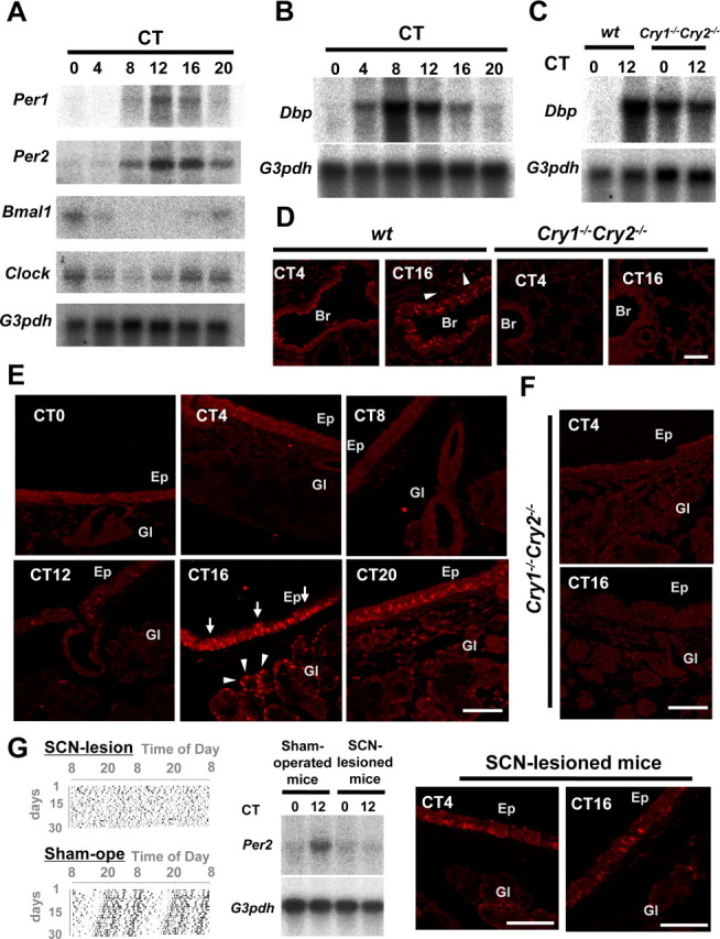
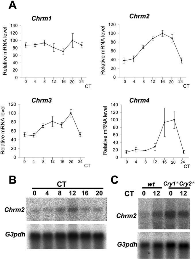
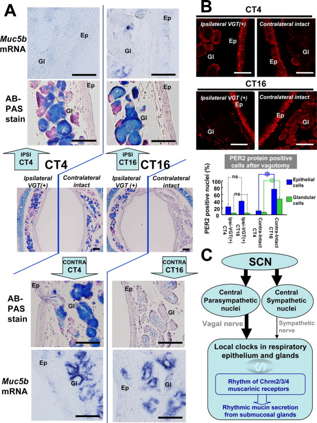
The present study addresses the role of the circadian system in day-night changes of respiratory functions in the mouse. In all airway tissues investigated (i.e., larynx, trachea, bronchus, and lung), we observed clear rhythmic expression of the Per1, Per2, Bmal1, and Clock core oscillator genes (the latter two genes oscillating in antiphase with the Per genes), as well as the clock-regulated Dbp gene. Oscillations were abolished in arrhythmic Cry1-/- Cry2-/- knock-out mice and after lesioning of the master clock in the suprachiasmatic nucleus (SCN) in wild-type animals. These findings indicate that respiratory system cells contain a functional peripheral oscillator that is controlled by the SCN. Furthermore, we found that the muscarinic acetylcholine receptor genes Chm2, Chm3, and Chm4 are expressed in a circadian manner, and that mucin secretion (rather than synthesis) by the airway submucosal glands is under circadian control. Signals from the SCN are mainly transmitted by the vagal nerve because unilateral vagotomy completely abolished rhythms in mucin and PER2 protein levels in the (operated) ipsilateral side of the submucosal glands, but not in the (intact) contralateral side. Thus, peripheral clock mediated circadian expression of muscarinic acetylcholine receptor proteins, and parasympathetic signaling between SCN and respiratory tissues are essential gears in conferring circadian "time" information to airway glands.


Figures

Figure 1.
Figure 1.
Clock genes and clock controlled genes in the mouse respiratory system. A, Circadian expression of Per1, Per2, Bmal1, and Clock genes, as detected by Northern blot analysis. G3pdh expression was determined as a control. Note the peak and through in Per1 and Per2 mRNA levels at CT12 and CT0, respectively, as well as the inverted rhythms of Bmal1 and Clock gene expression. B, Circadian expression of Dbp, a clock controlled gene. Dbp mRNA levels show a similar distribution pattern as that of Per1. C, Dbp mRNA levels in arrhythmic Cry1−/−Cry2−/− mice. D, Immunohistochemical staining of the PER2 protein in the lung and bronchiole of wild-type and Cry1−/−Cry2−/− mice at CT4 and CT16. PER2 proteins were abundantly expressed in the nucleus of bronchial epithelial cells at CT16, although almost absent at CT4. Alveolar cells (arrowheads) also showed a slight rhythm. PER2 protein expression was abolished in Cry1−/−Cry2−/− mice at any time examined. E, Circadian expression of the PER2 protein in epithelium and submucosal glands in trachea. Note the high level of nuclear immunohistochemical staining in tracheal epithelium (arrows) and submucosal glands (arrowheads) at CT16. F, Abolished PER2 protein expression in Cry1−/−Cry2−/− mice. G, Loss of circadian oscillation in Per2 mRNA and PER2 protein levels in the respiratory system of SCN-lesioned animals. Shown are (double-plotted) behavioral actograms (left), and Per2 mRNA expression levels at CT0 and CT12, as determined by Northern blot analysis (middle) of SCN-lesioned and sham-operated wild-type mice, as well as PER2-immunohistochemistry in SCN-lesioned mice at CT4 and CT16. Data were adopted as control versus sham-operated animals. Br, Bronchiole; Ep, tracheal epithelium; Gl, submucosal gland. Scale bars, 100 μm.
Figure 2.
Figure 2.
The circadian gene expression profiles of Chrm1, Chrm2, Chrm3, and Chrm4. A, Quantitative real-time PCR analysis of muscarinic acetylcholine receptor (Chrm1–4) gene expression in the lung of C57BL/6 mice at various circadian times. G3pdh expression was determined as a control. Whereas Chrm1 mRNA levels did not oscillate in a circadian manner, Chrm2, Chrm3, and Chrm4 transcription showed significant circadian rhythmicity, peaking at CT16–CT20. B, Northern blot analysis of Chrm2 mRNA levels in the lung of C57BL/6 mice, showing clear circadian rhythmicity. C, Northern blot analysis of Chrm2 expression in the lungs of Cry1−/−Cry2−/− mice and wild-type littermates at CT0 and CT12. Note the constant high Chrm2 mRNA levels in Cry1−/−Cry2−/− mice.
Figure 3.
Figure 3.
Vagotomy-induced changes in mucin levels and clock gene expression. A, Mucin levels (AB/PAS) and Muc5b gene expression (in situ hybridization) at CT4 and CT16 in the trachea of C57BL/6 mice, one week after hemivagotomy. Middle, Low-magnification photographs of the AB/PAS-stained trachea, showing the ipsilateral vagotomized [VGT(+)] side and the contralateral intact side. Top, bottom, High-magnification photographs of the AB/PAS-stained ipsilateral vagotomized side and contralateral intact side, respectively. In addition, Muc5b mRNA levels (as determined by in situ hybridization) at the ipsilateral side (top panels) and the contralateral intact side (bottom panels) are shown at high magnification. B, Tracheal PER2 protein after vagotomy at CT4 and CT16. Br, Bronchiole; Ep, tracheal epithelium; Gl, submucosal gland. The number of PER2 protein-positive nuclei in epithelial cells and glandular cells in trachea was calculated in the ipsilateral vagotomized side at CT4 (Ipsi-VGT(+)-CT4), ipsilateral vagotomized side at CT16 [Ipsi-VGT(+)-CT16], contralateral intact side at CT4 (Contra-Intact-CT4), and contralateral intact side at CT16 (Contra-Intact-CT16). Note the absence of rhythmic mPER2 expression at the vagotomized side, in contrast to the clear rhythm in contralateral intact side in both epithelial and glandular cells. Statistics was performed by Student's t test (mean ± SEM; n = 3; *p < 0.05). ns, Not significant. C, Model for the circadian control of mucin secretion in respiratory organs. Circadian expression of the muscarinic cholinergic receptor genes Chrm2, Chrm3, and Chrm4 is under control of the local peripheral circadian clock. The peripheral oscillator receives input from the central circadian oscillator in the SCN via the autonomous nervous system, in which vagal innervation is highly dominant over sympathetic innervation. Mucin production is constant, but the release of this glycoconjugate from the submucosal gland is increased at night time via increased muscarinic receptor expression and increased vagal tone. Scale bars: 100 μm.

References

    1. Barnes PJ. Circadian variation in airway function. Am J Med. 1985;79:5–9. - PubMed
    1. Barnes PJ. Muscarinic receptor subtypes in airways. Life Sci. 1993;52:521–527. - PubMed
    1. Buijs RM, Kalsbeek A. Hypothalamic integration of central and peripheral clocks. Nat Rev Neurosci. 2001;2:521–526. - PubMed
    1. Chen Y, Zhao YH, Wu R. In silico cloning of mouse Muc5b gene and upregulation of its expression in mouse asthma model. Am J Respir Cirt Care Med. 2001;164:1059–1066. - PubMed
    1. Damiola F, Minh N, Prenitner N, Kornmann B, Fleury-Olela F, Schibler U. Restricted feeding uncouples circadian oscillators in peripheral tissues from the central pacemaker in the suprachiasmatic nucleus. Genes Develop. 2000;14:2950–2961. - PMC - PubMed

Publication types

MeSH terms

LinkOut - more resources