. 2007 Apr 18;27(16):4443-51.
doi: 10.1523/JNEUROSCI.0557-07.2007.

Activation of TRPA1 channel facilitates excitatory synaptic transmission in substantia gelatinosa neurons of the adult rat spinal cord

Affiliations

Activation of TRPA1 channel facilitates excitatory synaptic transmission in substantia gelatinosa neurons of the adult rat spinal cord

Masafumi Kosugi et al. J Neurosci. .

Abstract

TRPA1 is expressed in primary sensory neurons and hair cells, and it is proposed to be activated by cold stimuli, mechanical stimuli, or pungent ingredients. However, its role in regulating synaptic transmission has never been documented yet. In the present study, we examined whether activation of the TRPA1 channels affects synaptic transmission in substantia gelatinosa (SG) neurons of adult rat spinal cord slices by using the whole-cell patch-clamp technique. A chief ingredient of mustard oil, allyl isothiocyanate (AITC), superfused for 2 min markedly increased the frequency and amplitude of spontaneous EPSCs (sEPSCs), which was accompanied by an inward current. Similar actions were produced by cinnamaldehyde and allicin. The AITC-induced increases in sEPSC frequency and amplitude were resistant to tetrodotoxin (TTX) and La3+, whereas being significantly reduced in extent in a Ca2+-free bath solution. In the presence of glutamate receptor antagonists CNQX and AP5, AITC did not generate any synaptic activities. The AITC-induced increases in sEPSC frequency and amplitude were reduced by ruthenium red, whereas being unaffected by capsazepine. AITC also increased the frequency and amplitude of spontaneous inhibitory postsynaptic currents; this AITC action was abolished in the presence of TTX or glutamate receptor antagonists. These results indicate that TRPA1 appears to be localized not only at presynaptic terminals on SG neurons to enhance glutamate release, but also in terminals of primary afferents innervating onto spinal inhibitory interneurons, which make synapses with SG neurons. This central modulation of sensory signals may be associated with physiological and pathological pain sensations.


Figures
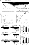
Figure 1.
Figure 1.
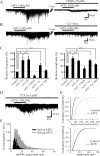
Actions of AITC and related substances on excitatory synaptic transmission in SG neurons. A, A continuous chart recording of glutamatergic sEPSCs before and during the action of AITC (100 μm; top). Three consecutive traces of sEPSCs are shown in an expanded scale in time, before (bottom left) and during the action of AITC (bottom right). Note a slow inward current that is accompanied by increases in sEPSC frequency and amplitude (top). B, Cumulative distributions of the interevent interval (left) and amplitude (right) of sEPSCs, before (dotted line) and during (continuous line) the action of AITC. AITC shifted the interevent interval and amplitude to a shorter and a larger one, respectively (p < 0.05; Kolmogorov–Smirnov test). Data in A and B were obtained from the same neuron. C, When AITC (100 μm) was applied repeatedly at 20 min intervals, it produced similar increases in sEPSC frequency and amplitude and induced an inward current having a similar amplitude (top). CA (100 μm) mostly increased sEPSC frequency and amplitude in a neuron in which AITC increased sEPSC frequency and amplitude (middle). Allicin (100 μm) also increased sEPSC frequency and amplitude in a neuron in which AITC increased sEPSC frequency and amplitude (bottom). D, Summary of sEPSC frequency (top) and amplitude (bottom) under the action of AITC (n = 129), CA (n = 5), and allicin (n = 5), relative to those in the control. Vertical lines accompanied by bars show SEM. Statistical significance between data shown by bars is indicated by an asterisk; *p < 0.05. The holding potential (VH) used was −70 mV.
Figure 2.
Figure 2.
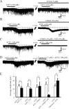
Characterization of the AITC-induced increases in sEPSC frequency and amplitude. A, Action of AITC (100 μm) on sEPSCs in the absence (left) and presence (right) of CNQX (10 μm). CNQX blocked sEPSCs not only in the absence of AITC but also under its action (right). B, Action of AITC on sEPSCs in normal Krebs' (left) and Ca2+-free solution (right). The AITC-induced increases in sEPSC frequency and amplitude were significantly reduced in extent in a Ca2+-free solution (right). C, Summary of sEPSC frequency (left) and amplitude (right) under the first [AITC (1), n = 10] and second application of AITC [AITC (2), n = 10], under the action of AITC in the presence of CNQX (n = 3) or La3+ (n = 5), and the action of AITC in Ca2+-free solution (n = 8), relative to those in the control. Vertical lines accompanied by bars show SEM. Statistical significance between data shown by bars is indicated by an asterisk; *p < 0.05. n.s., Not significant. D, A continuous chart recording of glutamatergic miniature EPSCs (mEPSCs) in the presence of TTX (0.5 μm) before and under the action of AITC (100 μm). E, Cumulative distributions of the interevent interval (top) and amplitude (bottom) of mEPSCs, before (dotted line) and during (continuous line) the action of AITC. AITC shifted the interevent interval and amplitude to a shorter and a larger one, respectively (p < 0.05; Kolmogorov–Smirnov test). F, Distributions of sEPSC amplitude before and during (filled bar and gray bar, respectively) the action of AITC. The bin width is 1 pA. VH = −70 mV.
Figure 3.
Figure 3.
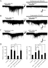
AITC induces a slow inward current in SG neurons. A, Action of AITC on the membrane holding current in the absence (left) and presence (right) of TTX (0.5 μm). The amplitude of the AITC-induced inward current did not change in the presence of TTX. B, Action of AITC on the membrane holding current in the absence (left) and presence (right) of CNQX (10 μm). In the presence of CNQX, AITC induced an inward current without any decrease in amplitude. C, Action of AITC on the membrane holding current in the absence (left) and presence (right) of CNQX (10 μm) and AP5 (50 μm). The addition of AP5 to CNQX significantly suppressed the AITC-induced inward current. D, When capsaicin (2 μm) was applied together with AITC (100 μm), the amplitude of the inward current was larger than that by AITC only. E, The average amplitudes of the inward currents induced by the first [AITC (1), n = 7] and second application of AITC in control [AITC (2), n = 7], and AITC in the absence and presence of TTX (n = 6), AITC in the absence and presence of CNQX (n = 5), AITC in the absence and presence of CNQX and AP5 (n = 5), and also by AITC (n = 5) only and both AITC and capsaicin (n = 5). VH = −70 mV.
Figure 4.
Figure 4.
Effects of TRP agonists and antagonists on sEPSC frequency and amplitude. A, Action of AITC (100 μm) on sEPSCs in the absence (left) and presence (right) of ruthenium red (300 μm). Ruthenium red significantly suppressed the AITC-induced increase in sEPSC frequency and amplitude. B, Action of AITC (100 μm) on sEPSCs in the absence (left) and presence (right) of capsazepine (10 μm). Capsazepine did not affect the AITC-induced increases in sEPSC frequency and amplitude. C, Capsaicin (2 μm) mostly increased sEPSC frequency and amplitude in a neuron. When AITC (100 μm) was applied at 20 min after washout of capsaicin, it still increased sEPSC frequency and amplitude and produced an inward current. D, Summary of sEPSC frequency (left) and amplitude (right) under the action of AITC (n = 129), AITC in the presence of capsazepine (n = 5), AITC in the presence of ruthenium red (n = 5), and AITC with the pretreatment of capsaicin (n = 5), relative to those in the control. Vertical lines accompanied by bars show SEM. Statistical significance between data shown by bars is indicated by an asterisk; *p < 0.05. n.s., Not significant. VH = −70 mV.
Figure 5.
Figure 5.
Action of AITC on inhibitory synaptic transmission in SG neurons. A, A continuous chart recording of sIPSCs before and during the action of AITC (100 μm). B, Cumulative distributions of the interevent interval (left) and amplitude (right) of sIPSCs, before (dotted line) and during the action of AITC (continuous line). AITC shifted the interevent interval and amplitude to a shorter and a larger one, respectively (p < 0.05; Kolmogorov–Smirnov test). C, A continuous chart recording of mIPSCs before and during the action of AITC (100 μm) in the presence of TTX (0.5 μm). TTX abolished the AITC-induced increases in sIPSC frequency and amplitude. D, A continuous chart recording of sIPSCs before and during the action of AITC in a mixture of CNQX (10 μm) and AP5 (50 μm). The AITC-induced increase in sIPSC frequency and amplitude was attenuated in the presence of the drugs. VH = 0 mV.
Figure 6.
Figure 6.
Schematic illustration of possible sites on which AITC acts in the SG. Schematic illustration of a proposed synaptic convergence of distinct TRPA1-sensitive sensory pathways onto SG neurons. Both central neurons and islet cells in the SG receive monosynaptic excitatory input from different primary afferent fibers. Central neurons receive an input from primary afferent fibers, which are sensitive to both TRPA1 and TRPV1, or TRPV1 only, whereas islet cells receive an input from TRPA1-sensitive and TRPV1-insensitive primary afferent fibers. The islet cell is an inhibitory interneuron and presynaptic to a central neuron.

Similar articles

Cited by

References

    1. Bandell M, Story GM, Hwang SW, Viswanath V, Eid SR, Petrus MJ, Earley TJ, Patapoutian A. Noxious cold ion channel TRPA1 is activated by pungent compounds and bradykinin. Neuron. 2004;41:849–857. - PubMed
    1. Bautista DM, Jordt S-E, Nikai T, Tsuruda PR, Read AJ, Poblete J, Yamoah EN, Basbaum AI, Julius D. TRPA1 mediates the inflammatory actions of environmental irritants and proalgesic agents. Cell. 2006;124:1269–1282. - PubMed
    1. Clapham DE. TRP channels as cellular sensors. Nature. 2003;426:517–524. - PubMed
    1. Corey DP, García-Añoveros J, Holt JR, Kwan KY, Lin S-Y, Vollrath MA, Amalfitano A, Cheung EL-M, Derfler BH, Duggan A, Géléoc GSG, Gray PA, Hoffman MP, Rehm HL, Tamasauskas D, Zhang D-S. TRPA1 is a candidate for the mechanosensitive transduction channel of vertebrate hair cells. Nature. 2004;432:723–730. - PubMed
    1. Gu JG, MacDermott AB. Activation of ATP P2X receptors elicits glutamate release from sensory neuron synapses. Nature. 1997;389:749–753. - PubMed

Publication types

LinkOut - more resources