. 2007 Jul;18(7):2389-99.
doi: 10.1091/mbc.e06-12-1128. Epub 2007 Apr 18.

MTOC reorientation occurs during FcgammaR-mediated phagocytosis in macrophages

Affiliations

MTOC reorientation occurs during FcgammaR-mediated phagocytosis in macrophages

Edward W Eng et al. Mol Biol Cell. 2007 Jul.

Abstract

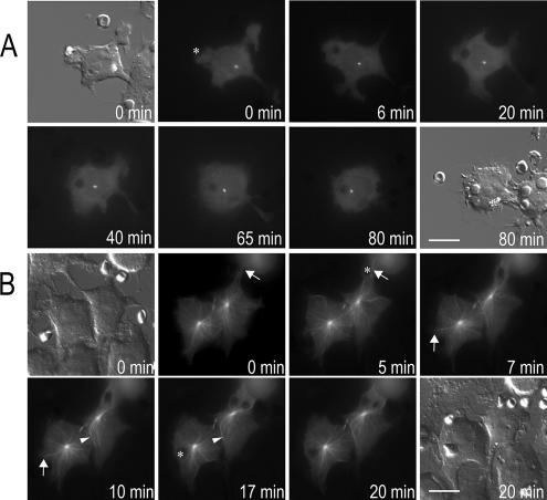
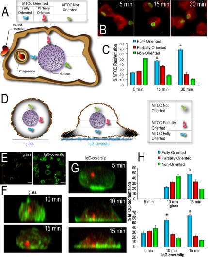
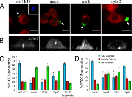
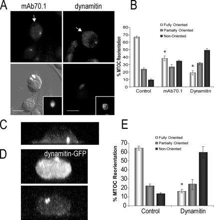
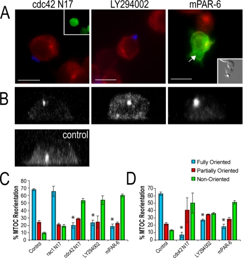
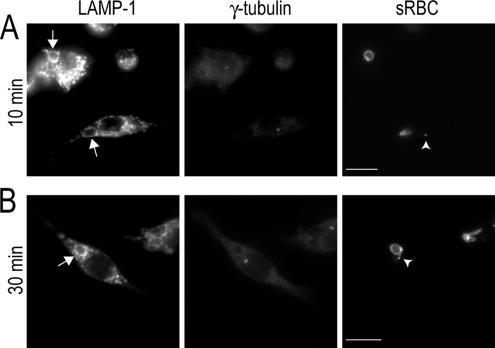
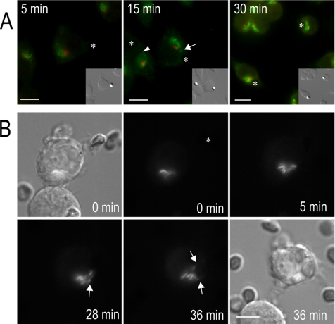
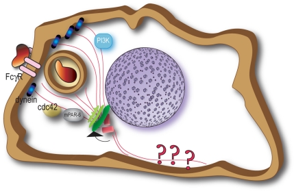
Cell polarization is essential for targeting signaling elements and organelles to active plasma membrane regions. In a few specialized cell types, cell polarity is enhanced by reorientation of the MTOC and associated organelles toward dynamic membrane sites. Phagocytosis is a highly polarized process whereby particles >0.5 microm are internalized at stimulated regions on the cell surface of macrophages. Here we provide detailed evidence that the MTOC reorients toward the site of particle internalization during phagocytosis. We visualized MTOC proximity to IgG-sRBCs in fixed RAW264.7 cells, during live cell imaging using fluorescent chimeras to label the MTOC and using frustrated phagocytosis assays. MTOC reorientation in macrophages is initiated by FcgammaR ligation and is complete within 1 h. Polarization of the MTOC toward the phagosome requires the MT cytoskeleton and dynein motor activity. cdc42, PI3K, and mPAR-6 are all important signaling molecules for MTOC reorientation during phagocytosis. MTOC reorientation was not essential for particle internalization or phagolysosome formation. However Golgi reorientation in concert with MTOC reorientation during phagocytosis implicates MTOC reorientation in antigen processing events in macrophages.


Figures

Figure 1.
Figure 1.
MTOC reorientation during phagocytosis in macrophages. (A and B) DIC and epifluorescent images of a RAW264.7 cell transfected with γ-tubulin-GFP (A) or CLIP-170-head-GFP (B). (See also Supplementary Movies 1A and 1B.) RAW264.7 cells were exposed to IgG-sRBCs, and time 0 represents time of sRBC attachment. Movement of γ-tubulin-GFP begins 6 min after sRBC contact (A) and directional MTOC movement toward the phagosome in CLIP-170-head-GFP–transfected cells starts at 5 min (B, cell on right) and 10 min (B, cell on left). Asterisk indicates the site of sRBC internalization in epifluorescent stills, arrows indicate MTs at the cell cortex at the site of phagocytosis, and arrowheads indicate the cortical MTs diagonal and posterior to the MTOC and phagosome. Scale bars, 10 μm.
Figure 2.
Figure 2.
Assays for MTOC reorientation during phagocytosis. (A) Cartoon illustration of MTOC reorientation during phagocytosis “zonal” scoring regime in immunostained cells, as described in Materials and Methods. Solid gray bars divide zones used to quantify MTOC reorientation during phagocytosis. (B) RAW264.7 cells were exposed to IgG-sRBCs and fixed at given time intervals. Cells were immunostained for γ-tubulin (red) and sRBC (green). (C) Quantification of MTOC reorientation during phagocytosis at the indicated time intervals. *p < 0.05 compared with 5 min. Data are mean ± SEM from three experiments (n > 100). (D) Illustration of MTOC reorientation scoring protocol for cells plated on glass or on IgG-coated coverslips to induce “frustrated phagocytosis” (see Materials and Methods). (E) Representative XY confocal images for RAW264.7 cells plated on glass or IgG-coated coverslips at 15 min and stained for γ-tubulin (red) and actin (green). (F and G) XZ confocal reconstructions of RAW264.7 cells plated on glass (F) or IgG-opsonized coverslips (G) for indicated time intervals. Cells were fixed and stained for γ-tubulin (red) and actin (green). (H) Quantification of MTOC reorientation in RAW264.7 cells plated on glass or IgG coverslips for indicated time intervals. *p < 0.05 compared with 10 min for glass and 5 min for IgG coverslips. Data are mean ± SEM from three experiments (n > 30). Scale bars, 10 μm.
Figure 3.
Figure 3.
MTs are required for MTOC reorientation during phagocytosis and actin is necessary for MTOC reorientation toward smaller particles. (A) Immunostaining of MTOC in RAW264.7 cells transfected with rac1 N17-GFP (blue, inset), or treated with 10 μM nocodazole, 10 μM colchicine, or 2 μM cytochalasin D. After 30 min of exposure to IgG-sRBCs or large 8-μm IgG-beads, cells were fixed and immunostained for γ-tubulin (red) and IgG-sRBCs (green). Cells transfected with rac1 N17-GFP served as a control for MTOC reorientation in cells where particle internalization is inhibited. Arrow indicates bound sRBC location on RAW264.7 cells. (B) XZ confocal reconstructions of untreated RAW264.7 cells and cells treated with MT and actin inhibitors, before plating on IgG coverslips for 15 min. (C) MTOC reorientation shown in A was quantified with respect to single, bound sRBCs in RAW264.7 cells, in addition to the quantification of MTOC reorientation in cytochalasin D–treated cells exposed to large 8-μm IgG-beads. *p < 0.05 compared with rac1 N17-GFP cells. Data are mean ± SEM from three replicate experiments (n > 30). (D) Quantification of MTOC reorientation during frustrated phagocytosis in control (untreated) cells, or nocodazole-, colchicine- or cytochalasin D–treated RAW264.7 cells. *p < 0.05 compared with control. Data are mean ± SEM from three replicate experiments (n > 30). Scale bars, 10 μm.
Figure 4.
Figure 4.
Dynein is essential for polarization of the MTOC during phagocytosis. (A) Immunofluorescence and DIC images of RAW264.7 cells microinjected with monoclonal anti-dynein (clone 70.1) antibodies or transfected with p50 dynamitin-GFP (insets show microinjected and transfected cell, respectively). Cells were exposed to IgG-beads or IgG-sRBCs for 30 min and fixed and immunostained for pericentrin and γ-tubulin, respectively (top panels). Before fixation, external sRBCs were lysed with water, and external beads were immunostained to differentiate between bound versus internalized particles. Arrows indicate location of internalized IgG-bead/sRBCs. (B) Quantification of MTOC reorientation toward the IgG-bead/sRBC from three replicate experiments. *p < 0.05 compared with control. Data are mean ± SEM from three experiments (n > 15). (C and D) XZ confocal reconstructions of nontransfected RAW264.7 cells (C) and cells transfected with p50 dynamitin-GFP (D) after plating on IgG coverslips for 15 min and immunostaining for γ-tubulin (C and D, bottom panel). (E) Quantification of MTOC reorientation during frustrated phagocytosis in control (nontransfected) cells or p50 dynamitin-GFP transfected RAW264.7 cells. *p < 0.05 compared with control. Data are mean ± SEM from three experiments (n > 15). Scale bars, 10 μm.
Figure 5.
Figure 5.
MTOC reorientation during phagocytosis requires cdc42, PI3K and mPAR-6. (A) Fluorescent images of RAW264.7 cells transfected with cdc42 N17-GFP (green, inset) or mPAR-6-FLAG (green) constructs, or treated with 100 μM LY294002. After 30 min of exposure to IgG-sRBCs or IgG-beads (DIC, inset), cells were fixed and immunostained for pericentrin (red, transfected cells) or γ-tubulin (red, nontransfected cells) and sRBCs (blue). Arrow indicates internalized IgG-bead location within the cell. (B) XZ confocal reconstructions of untreated RAW264.7 cells, or cells transfected with cdc42 N17-GFP or mPAR-6-FLAG, or treated with 100 μM LY294002 before plating on IgG coverslips for 15 min. (C) MTOC reorientation was quantified with respect to single, bound, or internalized IgG-sRBCs/beads in RAW264.7 cells. *p < 0.05 compared with control. Data are mean ± SEM from three replicate experiments (n > 30). (D) Quantification of MTOC reorientation during frustrated phagocytosis in control (untreated) cells, LY294002-treated, or cdc42 N17-GFP–, or mPAR-6-FLAG–transfected RAW264.7 cells. *p < 0.05 compared with control. Data are mean ± SEM from three replicate experiments (n > 30). Scale bars, 10 μm.
Figure 6.
Figure 6.
MTOC reorientation is not required for phagolysosome formation. Fluorescent images of RAW264.7 cells immunostained for LAMP-1 and γ-tubulin after 10 (A) and 30 min (B) of phagocytosis of IgG-sRBCs. Arrows indicate LAMP-1 accumulation around phagosomes. Arrowheads indicate sRBC fragments that are toward the MTOC, detected with anti-rabbit antibodies. Scale bars, 10 μm.
Figure 7.
Figure 7.
Golgi polarization occurs toward the phagosome. (A) RAW264.7 cells were exposed to IgG-beads (asterisk, and DIC image in insets) and fixed at given time intervals. Cells were immunostained for pericentrin (red) and giantin (green). Arrow indicates Golgi tubules extending toward the phagosome, and the arrowhead indicates Golgi tubules moving over the nucleus toward the phagosome. (B) Epifluorescent and DIC stills of a RAW264.7 cell transfected with luminal-GFP to label the Golgi, undergoing phagocytosis of an IgG-sRBC. Times are indicated from the time of particle contact. Asterisk marks initial site of bound sRBC. Scale bars, 10 μm.
Figure 8.
Figure 8.
A model of MTOC reorientation during FcγR-phagocytosis in macrophages. Shown in the illustration are the major receptor, cytoskeletal and intracellular signaling regulators of MTOC reorientation during phagocytosis. MTOC reorientation during phagocytosis is proposed to accelerate interactions of the phagosome with the Golgi complex.

Similar articles

Cited by

References

    1. Ackerman A. L., Kyritsis C., Tampe R., Cresswell P. Access of soluble antigens to the endoplasmic reticulum can explain cross-presentation by dendritic cells. Nat. Immunol. 2005;6:107–113. - PubMed
    1. Athlin L., Domellof L., Norberg B. O. The phagocytosis of yeast cells by monocytes: effects of colchicine, beta-lumicolchicine, and gamma-lumicolchicine. Lymphology. 1986;19:130–135. - PubMed
    1. Bergmann J. E., Kupfer A., Singer S. J. Membrane insertion at the leading edge of motile fibroblasts. Proc. Natl. Acad. Sci. USA. 1983;80:1367–1371. - PMC - PubMed
    1. Blocker A., Severin F. F., Burkhardt J. K., Bingham J. B., Yu H., Olivo J. C., Schroer T. A., Hyman A. A., Griffiths G. Molecular requirements for bi-directional movement of phagosomes along microtubules. J. Cell Biol. 1997;137:113–129. - PMC - PubMed
    1. Burkhardt J. K., Echeverri C. J., Nilsson T., Vallee R. B. Overexpression of the dynamitin (p50) subunit of the dynactin complex disrupts dynein-dependent maintenance of membrane organelle distribution. J. Cell Biol. 1997;139:469–484. - PMC - PubMed

Publication types

LinkOut - more resources