. 2007 Nov;56(11):1817-29.
doi: 10.1007/s00262-007-0325-0. Epub 2007 Apr 19.

DC immunotherapy is highly effective for the inhibition of tumor metastasis or recurrence, although it is not efficient for the eradication of established solid tumors

Affiliations

DC immunotherapy is highly effective for the inhibition of tumor metastasis or recurrence, although it is not efficient for the eradication of established solid tumors

Dae-Seog Lim et al. Cancer Immunol Immunother. 2007 Nov.

Abstract

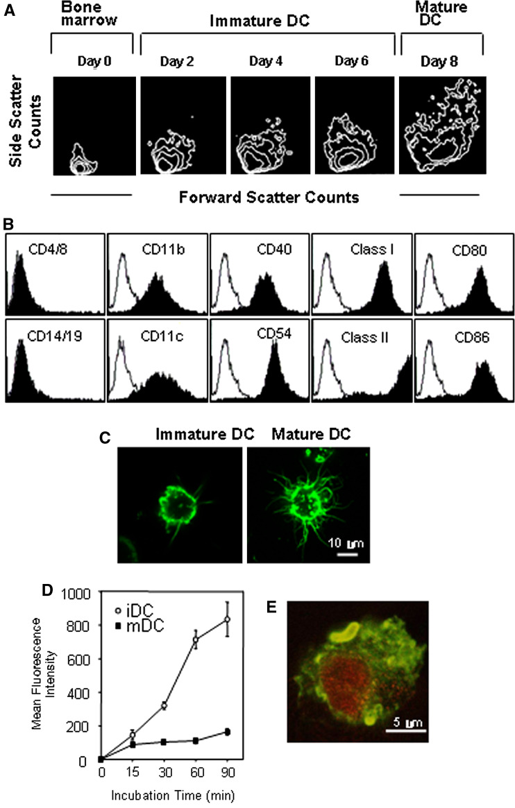
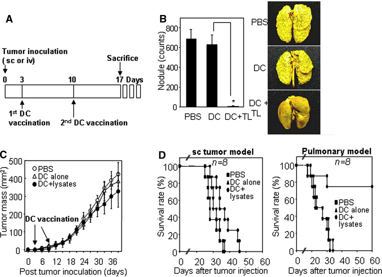
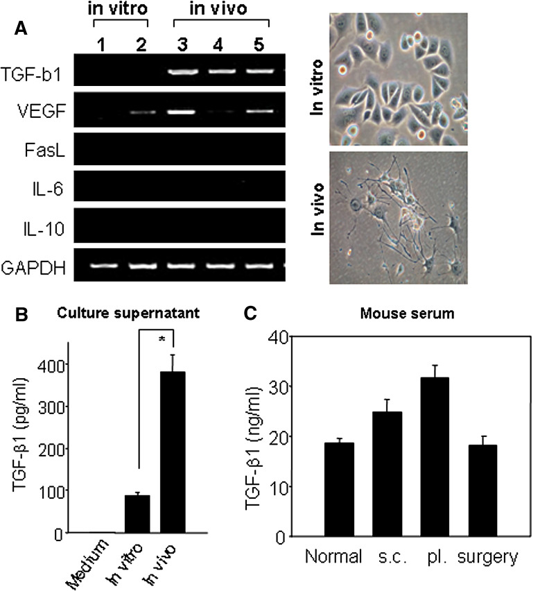
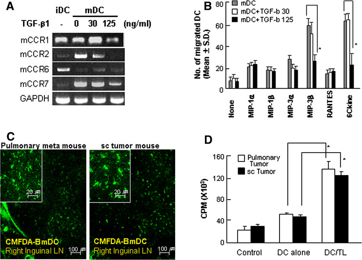
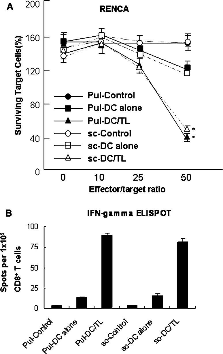
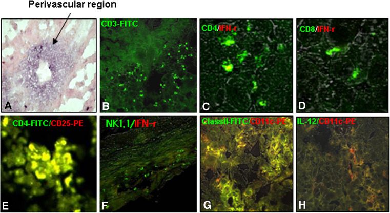
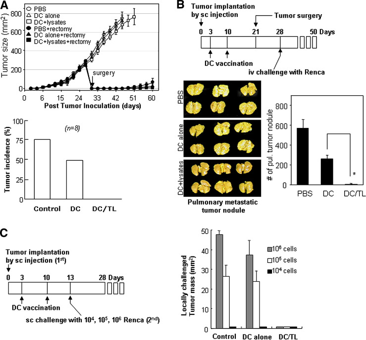
Dendritic cell (DC)-based immunotherapy has not been as effective as expected in most solid tumors even in the murine model, particularly in renal cell carcinoma (RCC). Our investigation was initiated to identify what causes the limitations of DC-based immunotherapy in solid RCC. We have investigated immunosuppressive factors from tumors and their effects on DC migration, as well as cytotoxic T lymphocyte (CTL) response and lymphocyte infiltration into the tumor mass upon vaccination with mouse renal adenocarcinoma (Renca) cell lysate-pulsed bone marrow (Bm)-derived DC in tumor-bearing mice. We also investigated pulmonary metastasis- and tumor recurrence-inhibitory effects of DC-vaccination in the solid tumor-bearing mice. In these experiments, we found that the limitations of DC-based immunotherapy to solid RCC likely result from tumor-mediated TGF-beta hindrance of immune attack rather than insufficient immune induction by DC therapy. In fact, the CTL response induced by DC therapy was quite sufficient and functional for the inhibition of tumor recurrence after surgery or of tumor metastasis induced by additional tumor-challenge to the tumor-bearing mice. Taken together, our present results obtained in mouse model suggest the potential of DC immunotherapy in tumor patients for hindering or blocking disease progression by inhibition of tumor metastasis and/or tumor recurrence after surgery.


Figures

Fig. 1
Fig. 1
Generation and characterization of BmDCs from Balb/c mice. a Increased cell size and granulity depend on the differential stages. b Surface phenotype of BmDC matured in the presence of INF-γ and TNF-α after tumor-lysate pulsing. Closed histograms show staining with test antibodies. c Representative morphology of immature (day 6) and mature (day 8) BmDCs. Cells were stained with FITC-conjugated anti-MHC class II antibody at the indicated time point. Representative images were obtained by confocal microscopy with a magnification of ×2,000 (software zoom ×2, original magnification ×1,000). d FITC-dextran uptake by immature and mature BmDCs. Cells were equilibrated at 37°C or 0°C prior to pulsing, and were then pulsed with fluorescein-conjugated dextran (40,000 m.w.) at a concentration of 1 mg/ml. After different durations of incubation, cold buffer was added to stop the reaction. Cells were analyzed using FACS, and non-specific binding was subtracted (incubation at 0°C). iDC immature DC, mDC mature DC. e Uptake of apoptotic Renca tumor cells by BmDCs (day 6). 7-AAD labeled apoptotic Renca tumor cells (red) were incubated with day 6-BmDCs (green), which were labeled with FITC-conjugated MHC class II antibody for 1 h. After washes with PBS, cells were visualized under a confocal spectral microscope with a magnification of ×4,000 (software zoom ×4, original magnification ×1,000)
Fig. 2
Fig. 2
DC vaccination was highly effective for survival and tumor regression in pulmonary metastatic tumor-bearing mice, but was not so effective for those of sc tumor-bearing mice. a A vaccination schedule. b Inhibition of pulmonary metastasis by vaccination with Renca tumor lysate-pulsed DCs. Balb/c mice received 1 × 106 viable Renca tumor cells in the lateral tail vein (i.v.) to establish pulmonary metastasis. Mice were then inoculated sc in the right flank with 1 × 106 DCs (Renca tumor lysate-pulsed) twice on days 3 and 10 after tumor injection, and were euthanized on day 17. Tumor nodules on the lungs were visualized by staining with Bouin’s fixative for 10 min (right image). Tumor nodules (shown in right panel) were enumerated, and the values represent the mean number of nodules ± SD of twenty mice per group (left panel). DC + TL tumor lysate-pulsed DC. Asterisk P < 0.01. PBS PBS control, DC alone mature DC, DC + lysates tumor lysate-pulsed DC. c Sc tumor progression after immunization with Renca tumor lysate-pulsed DC. Mice were inoculated (sc) with 1 × 106 viable Renca tumor cells in the left flank and immunized (sc) with Renca tumor lysate-pulsed DC in the right flank, as described above. The size of the tumor mass was recorded every 3 days as the tumor area (in square mm), and was determined by measuring the largest perpendicular diameter with calipers. Data shown are representative of five experiments and are reported as the average tumor size ± SD of eight mice per group. d Survival percentage of both pulmonary metastatic tumor-bearing mice and sc tumor-bearing mice after DC vaccination. Mice were evaluated daily until death. Data are representative of at least two independent experiments with eight mice per group
Fig. 3
Fig. 3
TGF-β1 was enhanced for its expression among the immunosuppressive agents in Renca cells isolated from the tumor mass, but was not detected in Renca cells subcultured in vitro. a Screening of the expression of immunosuppressive agents by RT-PCR and their morphology from in vitro-cultured Renca tumor cells or in vivo-passaged tumor cells (subcutaneous). First-strand cDNA was synthesized from total RNA using reverse transcriptase and amplified with primers specific for TGF-β1, VEGF, Fas ligand, IL-6, IL-10 and GAPDH. Phase-contrast photographs were taken directly from the day 7 cultures of in vivo-passaged Renca tumor cells. Original magnification is ×400. 1, 2 In vitro-passaged Renca cells; 3, 4, 5 Renca cells obtained from three different tumor-bearing mice without any treatment, vaccinated with tumor lysate-pulsed DC, and treated with unpulsed DC, respectively. b Quantification of TGF-β1 in culture supernatants. Culture supernatants were prepared from 7-day cultures of Renca cells (1 × 106 cells) from sc tumor masses or in vitro subcultures. ELISA was performed as described in protocol of the manufacturer. Values shown are means ± SD of two experiments. Asterisk P < 0.01. In vitro in vitro-passaged tumor cells, in vivo cells from sc tumor. c Quantification of TGF-β1 in mouse sera. Mouse sera were obtained from sc tumor-bearing mice (s.c.), pulmonary metastatic tumor-bearing mice (pl.) on day 17 after tumor inoculation. The serum of tumor-rectomized mice (surgery) was prepared a week after surgery of day 17 sc tumor-bearing mice. Normal serum (normal) was prepared from 8-week-old Balb/c mice. ELISA was performed as described in protocol of the manufacturer. Values shown are means ± SD of 3 to 5 mice per group
Fig. 4
Fig. 4
No difference in the capacity of DC migration and T cell proliferation after DC vaccination between the mouse groups bearing pulmonary metastatic tumors and sc tumors, respectively. a Effect of TGF-β on the chemokine expression of mature DCs. Renca tumor lysate-pulsed DCs were incubated in the presence of 30 or 125 ng/ml of TGF-β1 for 24 h. First-strand cDNA was synthesized and amplified with primers for mCCR1, mCCR2, mCCR6, mCCR7, and GAPDH. iDC immature DC, mDC mature DC. b Effect of TGF-β on DC migration in chemotactic assay. Mature DCs were resuspended in RPMI-10 and treated with TGF-β. Chemokines at 1 μg/ml were placed in the lower chamber, and a filter with 5-μm pore size was placed on top. An aliquot of 1 × 106 cells/well were applied to the top surface of the filter, and the plate was incubated at 37°C for 4 h. Migrated cells were analyzed for CD11c expression by FACS. Each assay was performed in triplicate, and the results were expressed as the mean number of cells that migrate to the lower chamber ± SD. Asterisk P < 0.01. mDC mature DCs, mDC + TGF-b 30 mature DCs treated with 30 ng/ml TGF-β, mDC + TGF-b 125 mature DCs treated with 125 ng/ml TGF-β. c Immunohistochemistry of DC migration into regional lymph nodes in pulmonary metastatic tumor- and sc tumor-bearing mice. Renca tumor lysate-pulsed DCs were labeled with CMFDA and intradermally injected into the left foot pads of the mice. After 18 h, inguinal lymph nodes were isolated from each immunized mouse (on day 17 after tumor implantation). Frozen tissues were sectioned at a thickness of 25 μm and mounted. The sections were then analyzed by confocal microscopy at a magnification of × 400 (inserted images). LN lymph node, Pulmonary meta mouse metastatic tumor-bearing mice, sc tumor mouse sc tumor-bearing mice. d Proliferation of T cells from sc or pulmonary metastatic tumor-bearing mice in response to Renca tumor lysate-pulsed DCs. Spleens were prepared from mice that were immunized as described in “Materials and methods” and stimulated with Renca tumor lysate-pulsed DCs for 5 days. Then, 1 μCi of tritiated deoxythymidine was added to each well for an additional 16 h. Incorporation of thymidine was determined using a liquid scintillation counter (see "Materials and methods"). Data shown are representative of two independent experiments. Asterisk P < 0.01. Control not stimulated, DC alone stimulated with mature DCs, DC/TL stimulated with Renca tumor lysate-pulsed DCs, Pulmonary tumor metastatic tumor-bearing mice, sc tumor sc tumor-bearing mice
Fig. 5
Fig. 5
DC vaccination induced similar intensities of tumor-specific CTLs and an increase of IFN-γ-secreting CD8 + T cells in both pulmonary metastatic tumor- and sc tumor-bearing mouse groups. a The induction of effective CTL responses by vaccination with Renca tumor lysate-pulsed DCs. CTLs were prepared by stimulation with Renca tumor lysate-pulsed DCs for 5 days and incubated with Renca tumor cells as a target for 24 h. After washing, target cells were stained with crystal violet and lysed using methanol. Surviving target cells were analyzed by measuring the absorbance at 470 nm. Data shown were representative of two independent experiments. Asterisk P < 0.01 compared with the control. b In vivo induction of IFN-γ-secreting CD8+ T cell responses. CD8+ T cells were isolated from post-vaccination spleen samples of mice that received the vaccination with Renca lysate-pulsed DCs (2 cycles of 1 × 106 cells per treatment). Cells were stimulated for 20 h with Renca lysate-pulsed DCs. IFN-γ-expressing T cells were enumerated using an automated ELISPOT reader, and activated T cell frequencies were expressed as the number of spot-forming cells per 1 × 105 CD8+ T cells. Pul-Control: unstimulated CTLs generated from metastatic tumor-bearing mice, Pul-DC alone mature DC-stimulated CTLs generated from metastatic tumor-bearing mice, Pul-DC/TL Renca tumor lysate-pulsed DC-stimulated CTLs generated from metastatic tumor-bearing mice, sc-control unstimulated CTLs generated from sc tumor-bearing mice, sc-DC alone mature DC-stimulated CTLs generated from sc tumor-bearing mice, sc-DC/TL Renca tumor lysate-pulsed DC-stimulated CTLs generated from sc tumor-bearing mice
Fig. 6
Fig. 6
TGF-β was clearly observed in sc tumor masses, and effector T cells were rarely observed. CD4+CD25+ Treg cells, inactivated T and NK cells, and immature DCs comprised the major population in the TILs. a TGF-β1 was highly concentrated around the perivascular regions in tumor masses (black arrow). bh Confocal immunohistochemistry. b Total T cells, c IFN-γ-producing CD4 T cells, d IFN-γ-producing CD8 T cells, e CD4+CD25+ Treg cells, f IFN-γ-producing NK/NKT cells, g MHC class II/CD11c-postive DCs, h IL-12-secreting DCs. See “Materials and methods” for the experimental procedures
Fig. 7
Fig. 7
DC vaccination was sufficient to inhibit further spread of metastatic tumors or tumor recurrence after surgery. a The upper panel shows inhibition of tumor recurrence after surgery in DC-vaccinated mice. The 3-day sc tumor-bearing mice were vaccinated twice, at a 1-week interval, with tumor lysate-pulsed DCs or unpulsed DCs. On day 30, the primary sc tumor of each mouse was surgically removed, and tumor recurrence was then assessed every three days. Data are reported as the average tumor area ± SD of eight mice per group. The lower panel shows the tumor recurrence rate after surgery in three different groups of mice. Control PBS-vaccinated group, DC alone mature DC alone-vaccinated group, DC/TL Renca tumor lysate-pulsed DC-vaccinated group. See “Materials and methods” for the experimental procedures. b Upper panel A schedule for DC vaccination, tumor surgery and tumor re-challenge in the sc tumor-bearing mice. Lower panels Pulmonary metastasis was inhibited in the tumor re-challenge experiment in DC-vaccinated sc tumor-bearing mice. Data are reported as the mean number of tumor nodules ± SD of six mice per group. Asterisk P < 0.01. PBS unvaccinated, DC mature DC-vaccinated, DC/TL Renca tumor lysate-pulsed DC-vaccinated. c Inhibition of sc tumors in the tumor re-challenge experiment with three different doses (left panel an experimental schedule). Data are reported as the average tumor area ± SD of four mice per group (right panel). Three different doses of Renca cells were used for re-challenge (sc) at the abdominal regions of the mice. Control PBS-vaccinated, DC alone mature DC-vaccinated, DC/TL Renca tumor lysate-pulsed DC-vaccinated. Data shown above are representative of two experiments

Similar articles

Cited by

References

    1. Ahn JH, Lee Y, Jeon C, Lee SJ, Lee BH, Choi KD, Bae YS. Identification of the genes differentially expressed in human dendritic cell subsets by cDNA subtraction and microarray analysis. Blood. 2002;100:1742–1754. - PubMed
    1. Amoils KD, Bezwoda WR. TGF-beta 1 mRNA expression in clinical breast cancer and its relationship to ER mRNA expression. Breast Cancer Res Treat. 1997;42:95–101. doi: 10.1023/A:1005785421815. - DOI - PubMed
    1. Arlen PM, Gulley JL. Therapeutic vaccines for prostate cancer: a review of clinical data. Curr Opin Investig Drugs. 2005;6:592–596. - PubMed
    1. Asselin-Paturel C, Echchakir H, Carayol G, Gay F, Opolon P, Grunenwald D, Chouaib S, Mami-Chouaib F. Quantitative analysis of Th1, Th2 and TGF-beta1 cytokine expression in tumor, TIL and PBL of non-small cell lung cancer patients. Int J Cancer. 1998;77:7–12. doi: 10.1002/(SICI)1097-0215(19980703)77:1<7::AID-IJC2>3.0.CO;2-Y. - DOI - PubMed
    1. Becker C, Fantini MC, Neurath MF. TGF-beta as a T cell regulator in colitis and colon cancer. Cytokine Growth Factor Rev. 2006;17:97–106. doi: 10.1016/j.cytogfr.2005.09.004. - DOI - PubMed

MeSH terms

Substances