

Renal cell carcinoma presenting with paraneoplastic hypercalcemic coma: a case report and review of the literature
- PMID: 17443359
- PMCID: PMC2219737
- DOI: 10.1007/s11606-007-0189-1
Renal cell carcinoma presenting with paraneoplastic hypercalcemic coma: a case report and review of the literature
Abstract
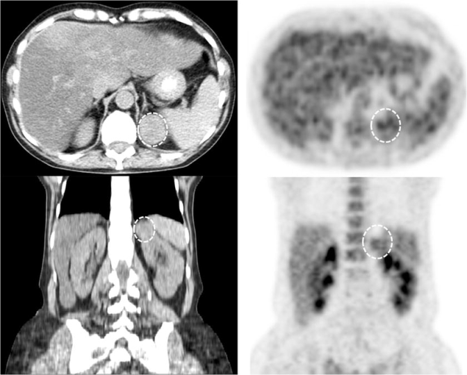
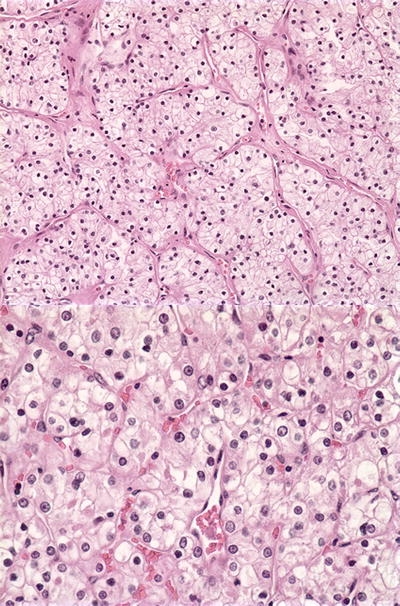
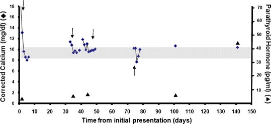
We report a case of a 62-year-old woman with renal cell carcinoma (RCC) presenting with a hypercalcemia-induced coma. A laboratory evaluation indicated nonparathyroid-mediated hypercalcemia with an initial serum calcium level of 18.6 mg/dL. Our patient's parathyroid hormone (PTH)-related peptide level was undetectable. Initial imaging was negative, but PET scan identified a mass in the upper pole of the left kidney. Our patient underwent partial nephrectomy, and the mass was identified as RCC on final pathology. After surgery, her hypercalcemia resolved and PTH returned to normal limits. This case report describes a patient with RCC with the unusual presentation of hypercalcemic coma. We review the differential diagnosis of malignant hypercalcemia and the evaluation of hypercalcemia occurring with RCC. This case illustrates the need to carefully review and interpret all available data, especially when conventional testing in the work-up of hypercalcemia is unrevealing.
Figures
References
-
- McLaughlin J, Lipworth L, Tarone R. Epidemiologic aspects of renal cell carcinoma. Semin Oncol. 2006;33(5):527–33. - PubMed
-
- Chow W, Devesa S, Warren J, et al. Rising incidence of renal cell cancer in the United States. JAMA. 1999;281(17):1628–31. - PubMed
-
- Cohen H, McGovern F. Renal-cell carcinoma. N Engl J Med. 2005;353(23)2477–90. - PubMed
-
- Zagoria R, Dyer R, Wolfman N, Hinn G, Chen Y. Radiology in the diagnosis and staging of renal cell carcinoma. Crit Rev Diagn Imaging. 1990;31(1):81–115. - PubMed
-
- Gutzeit A, Antoch G, Kuhl H, et al. Unknown primary tumors: detection with dual-modality PET/CT—initial experience. Radiology. 2005;234(1):227–34. - PubMed
Publication types
MeSH terms
Substances
LinkOut - more resources
Full Text Sources
Medical
