

Quantitative assessment of gestational sac shape: the gestational sac shape score
- PMID: 17444551
- PMCID: PMC3516405
- DOI: 10.1002/uog.3994
Quantitative assessment of gestational sac shape: the gestational sac shape score
Erratum in
- Ultrasound Obstet Gynecol. 2008 Oct;32(5):720
Abstract
Objective: To develop a quantitative method for characterizing gestational sac shape.
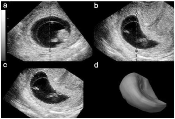
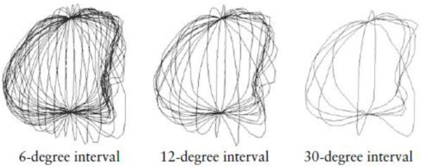
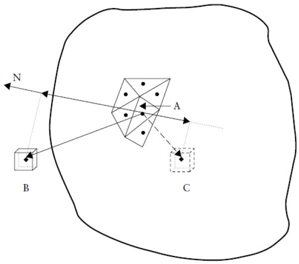
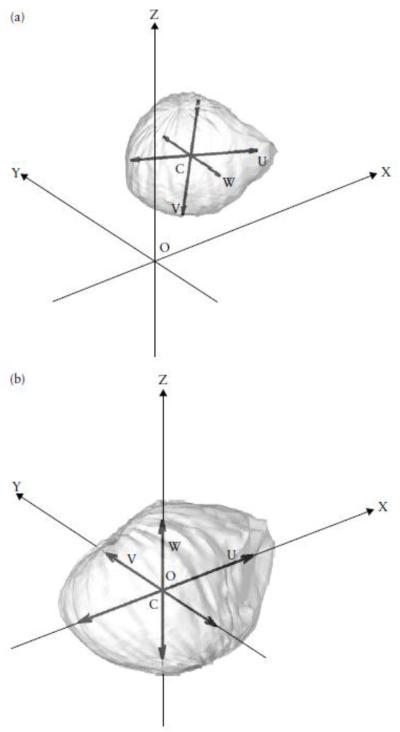
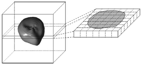
Methods: Twenty first-trimester gestational sacs in normal pregnancies were studied with three-dimensional (3D) ultrasonography. The 3D coordinates of surface-point sets were obtained for each sac using 30-, 15- and six-slice sampling. Cubic spline interpolation was used with the 15- and six-slice surface-point samples to generate coordinates for those 30-slice surface points not measured. Interpolated and measured values, the latter from the 30-slice sample, were compared and the percent error calculated. Cubic spline interpolation was used to determine the coordinates of a standard surface-point sample (3660) for each sac in each slice sample. These coordinate data were used to give each sac a standard configuration by moving its center of gravity to the origin, aligning its inertial axes along the coordinate axes and converting its volume to 1.0 mL. In this form, a volume shape descriptor could be generated for each sac that was then transformed into a vector containing only shape information. The 20 shape vectors of each slice sample were subjected to principal components analysis, and principal component scores (PCSs) calculated. The first four PCSs were used to define a gestational sac shape score (GSSS-30, GSSS-15 or GSSS-6) for each sac in a given slice sample. The characteristics of each set of GSSSs were determined and those for the GSSS-15 and GSSS-6 were compared with the GSSS-30 characteristics.
Results: Cubic spline interpolations were very accurate in most cases, with means close to 0%, and approximately 95% of the errors being less than 10%. GSSS-30 accounted for 67.6% of the shape variance, had a mean of zero and an SD of 1.1, was normally distributed and was not related to menstrual age (R=-0.16, P=0.51). GSSS-15 and GSSS-6 had essentially the same characteristics. No significant differences between individual GSSS-30 values and those for GSSS-15 or GSSS-6 were found, indicating the absence of a slice sample effect.
Conclusion: Using sophisticated mathematical methods, the gestational sac shape, initially represented by the 3D coordinates of 3660 surface points, was converted to a single number, the GSSS. This score had the appropriate properties for quantitatively characterizing normal, first-trimester gestational sac shapes. As it can be obtained from as few as six slices, it should be useful in many clinical situations. This novel approach has the potential for providing quantitative shape information about a variety of biological shapes and how they change over time.
Copyright (c) 2007 ISUOG.
Figures
Similar articles
-
Quantitative and morphological assessment of early gestational sacs using three-dimensional ultrasonography.Ultrasound Obstet Gynecol. 2006 Sep;28(3):255-60. doi: 10.1002/uog.2840. Ultrasound Obstet Gynecol. 2006. PMID: 16937412
-
Three-dimensional sonographic volumetry of the gestational sac and the amniotic sac in the first trimester.J Ultrasound Med. 2008 Mar;27(3):373-8. doi: 10.7863/jum.2008.27.3.373. J Ultrasound Med. 2008. PMID: 18314515
-
Sonography during early pregnancy: dependence of threshold and discriminatory values on transvaginal transducer frequency.AJR Am J Roentgenol. 1999 Apr;172(4):983-8. doi: 10.2214/ajr.172.4.10587132. AJR Am J Roentgenol. 1999. PMID: 10587132
-
Folic acid supplementation and malaria susceptibility and severity among people taking antifolate antimalarial drugs in endemic areas.Cochrane Database Syst Rev. 2022 Feb 1;2(2022):CD014217. doi: 10.1002/14651858.CD014217. Cochrane Database Syst Rev. 2022. PMID: 36321557 Free PMC article.
-
Sonographic signs of early pregnancy.Crit Rev Diagn Imaging. 1988;28(3):181-211. Crit Rev Diagn Imaging. 1988. PMID: 3044695 Review.
Cited by
-
Recognition of Fetal Facial Ultrasound Standard Plane Based on Texture Feature Fusion.Comput Math Methods Med. 2021 Jun 3;2021:6656942. doi: 10.1155/2021/6656942. eCollection 2021. Comput Math Methods Med. 2021. PMID: 34188691 Free PMC article.
References
-
- Lee W, Deter RL, McNie B, Powell M, Goncalves LF, Espinoza J, Romero R. Quantitative and morphological assessment of early gestational sacs using three-dimensional ultrasonography. Ultrasound Obstet Gynecol. 2006;28:255–260. - PubMed
-
- Metzenbauer M, Hafner E, Hoefinger D, Schuchter K, Stangl G, Ogris E, Philipp K. Three-dimensional ultrasound measurement of the placental volume in early pregnancy: method and correlation with biochemical placenta parameters. Placenta. 2001;22:602–605. - PubMed
-
- Goncalves LF, Lee W, Espinoza J, Romero R. Examination of the fetal heart by four-dimensional (4D) ultrasound with spatiotemporal image correlation (STIC) Ultrasound Obstet Gynecol. 2006;27:336–348. - PubMed
-
- Chang C-H, Yu C-H, Chang F-M, Ko H-C, Chen H-Y. The assessment of normal fetal liver volume by three-dimensional ultrasound. Ultrasound Med Biol. 2003;29:1123–1129. - PubMed
-
- Gerards FA, Engels AJ, Twisk JWR, Van Vugt JMG. Normal fetal lung volume measured with three-dimensional ultrasound. Ultrasound Obstet Gynecol. 2006;27:134–144. - PubMed
MeSH terms
Grants and funding
LinkOut - more resources
Full Text Sources
Research Materials
