

Effect of cell size and shape on single-cell electroporation
- PMID: 17444611
- PMCID: PMC2532982
- DOI: 10.1021/ac062049e
Effect of cell size and shape on single-cell electroporation
Abstract
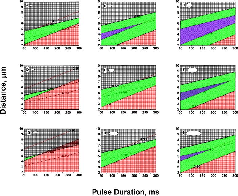
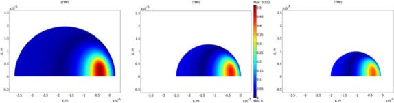
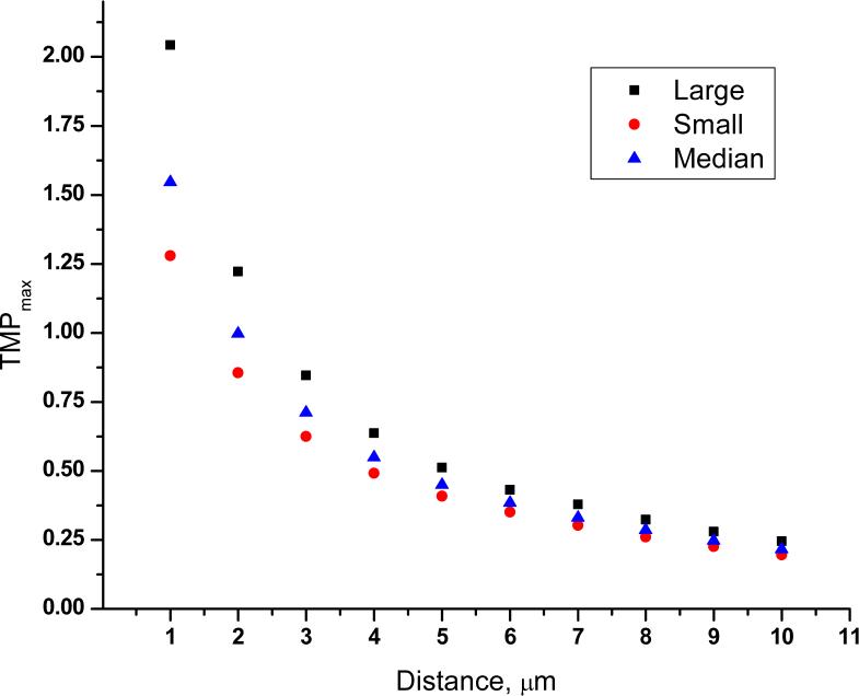
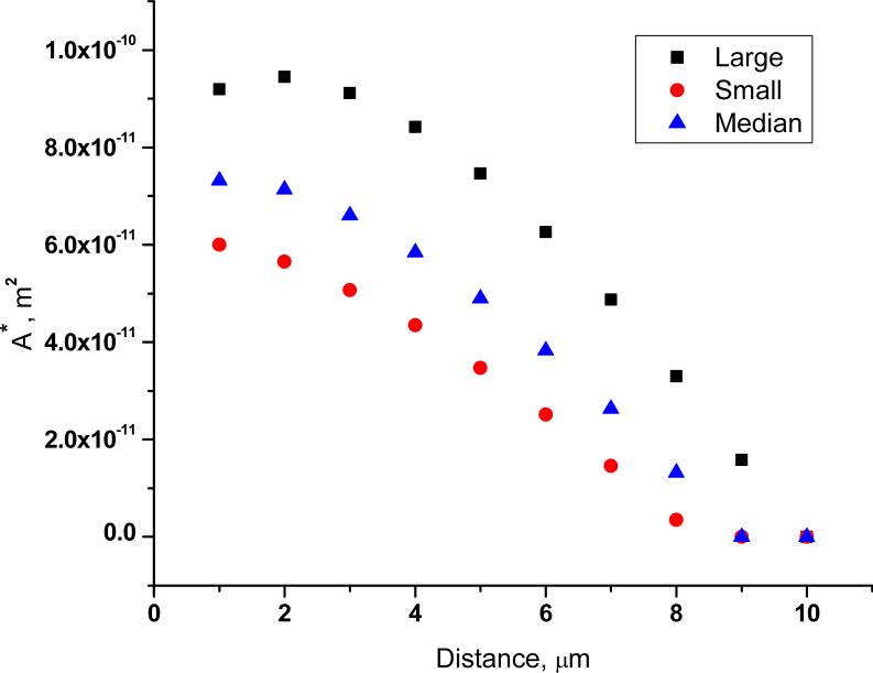
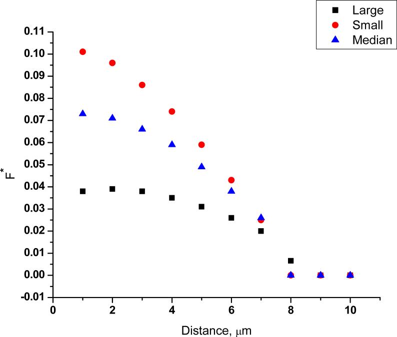
Single-cell electroporation was performed using electrolyte-filled capillaries on fluorescently labeled A549 cells. Cells were exposed to brief pulses (50-300 ms) at various cell-capillary tip distances. Cell viability and electroporation success were measured. In order to understand the variability in single-cell electroporation, logistic regression was used to determine whether the probabilities of cell survival and electroporation depend on experimental conditions and cell properties. Both experimental conditions and cell properties (size and shape) have a significant effect on the outcome. Finite element simulations were used to compare bulk electroporation to single-cell electroporation in terms of cell size and shape. Cells are more readily permeabilized and are more likely to survive if they are large and hemispherical as opposed to small and ellipsoidal with a high aspect ratio. The dependence of the maximum transmembrane potential across the cell membrane on cell size is much weaker than it is for bulk electroporation. Observed survival probabilities are related to the calculated fraction of the cell's surface area that is electroporated. Observed success of electroporation is related to the maximum transmembrane potential achieved.
Figures

Similar articles
-
Simultaneous maximization of cell permeabilization and viability in single-cell electroporation using an electrolyte-filled capillary.Anal Chem. 2007 Jan 1;79(1):161-7. doi: 10.1021/ac061270o. Anal Chem. 2007. PMID: 17194134 Free PMC article.
-
Numerical calculations of single-cell electroporation with an electrolyte-filled capillary.Biophys J. 2007 May 15;92(10):3696-705. doi: 10.1529/biophysj.106.097683. Epub 2007 Mar 9. Biophys J. 2007. PMID: 17351001 Free PMC article.
-
Scanning electroporation of selected areas of adherent cell cultures.Anal Chem. 2007 Jun 15;79(12):4410-8. doi: 10.1021/ac062140i. Epub 2007 May 19. Anal Chem. 2007. PMID: 17511419
-
Induced transmembrane voltage and its correlation with electroporation-mediated molecular transport.J Membr Biol. 2010 Jul;236(1):3-13. doi: 10.1007/s00232-010-9279-9. Epub 2010 Jul 9. J Membr Biol. 2010. PMID: 20617432 Review.
-
Chemical Enhancement of Irreversible Electroporation: A Review and Future Suggestions.Technol Cancer Res Treat. 2019 Jan 1;18:1533033819874128. doi: 10.1177/1533033819874128. Technol Cancer Res Treat. 2019. PMID: 31500518 Free PMC article. Review.
Cited by
-
Human cardiomyocytes are more susceptible to irreversible electroporation by pulsed electric field than human esophageal cells.Physiol Rep. 2022 Oct;10(20):e15493. doi: 10.14814/phy2.15493. Physiol Rep. 2022. PMID: 36301726 Free PMC article.
-
Physical and Chemical Enhancement of and Adaptive Resistance to Irreversible Electroporation of Pancreatic Cancer.Ann Biomed Eng. 2018 Jan;46(1):25-36. doi: 10.1007/s10439-017-1932-3. Epub 2017 Oct 5. Ann Biomed Eng. 2018. PMID: 28983745 Free PMC article.
-
Enhancing Irreversible Electroporation by Manipulating Cellular Biophysics with a Molecular Adjuvant.Biophys J. 2017 Jul 25;113(2):472-480. doi: 10.1016/j.bpj.2017.06.014. Biophys J. 2017. PMID: 28746857 Free PMC article.
-
In Vitro Electrochemistry of Biological Systems.Annu Rev Anal Chem (Palo Alto Calif). 2008 Jul 1;1:329. doi: 10.1146/annurev.anchem.1.031207.113038. Annu Rev Anal Chem (Palo Alto Calif). 2008. PMID: 20151038 Free PMC article.
-
The Emerging Landscape for Combating Resistance Associated with Energy-Based Therapies via Nanomedicine.Adv Mater. 2024 Feb;36(5):e2308286. doi: 10.1002/adma.202308286. Epub 2023 Nov 28. Adv Mater. 2024. PMID: 37971203 Free PMC article. Review.
References
-
- Neumann E, Kakorin S, Toensing K. Bioelectrochemistry and Bioenergetics. 1999;48:3–16. - PubMed
-
- Faurie C, Golzio M, Phez E, Teissie J, Rols MP. Engineering in Life Sciences. 2005;5:179–186.
-
- Bonnafous P, Vernhes MC, Teissie J, Gabriel B. Biochimica et Biophysica Acta, Biomembranes. 1999;1461:123–134. - PubMed
-
- Loste F, Eynard N, Teissie J. Bioelectrochemistry and Bioenergetics. 1998;47:119–127.
-
- Gift EA, Weaver JC. Cytometry. 2000;39:243–249. - PubMed
Publication types
MeSH terms
Grants and funding
LinkOut - more resources
Full Text Sources
Other Literature Sources
