. 2007 May 15;46(19):5754-65.
doi: 10.1021/bi602580c. Epub 2007 Apr 20.

Site-specific S-glutathiolation of mitochondrial NADH ubiquinone reductase

Affiliations

Site-specific S-glutathiolation of mitochondrial NADH ubiquinone reductase

Chwen-Lih Chen et al. Biochemistry. .

Abstract

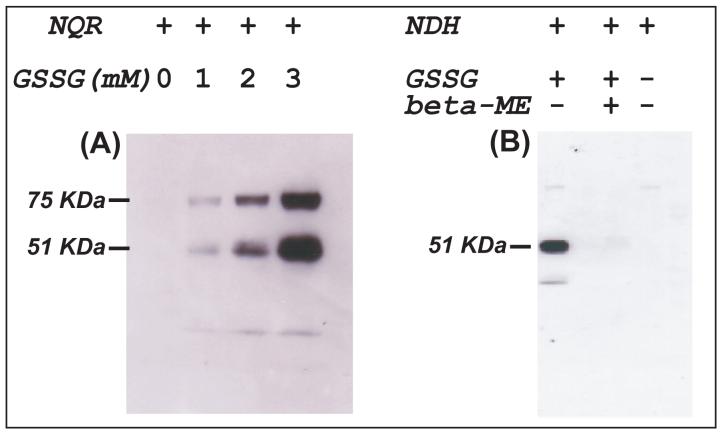
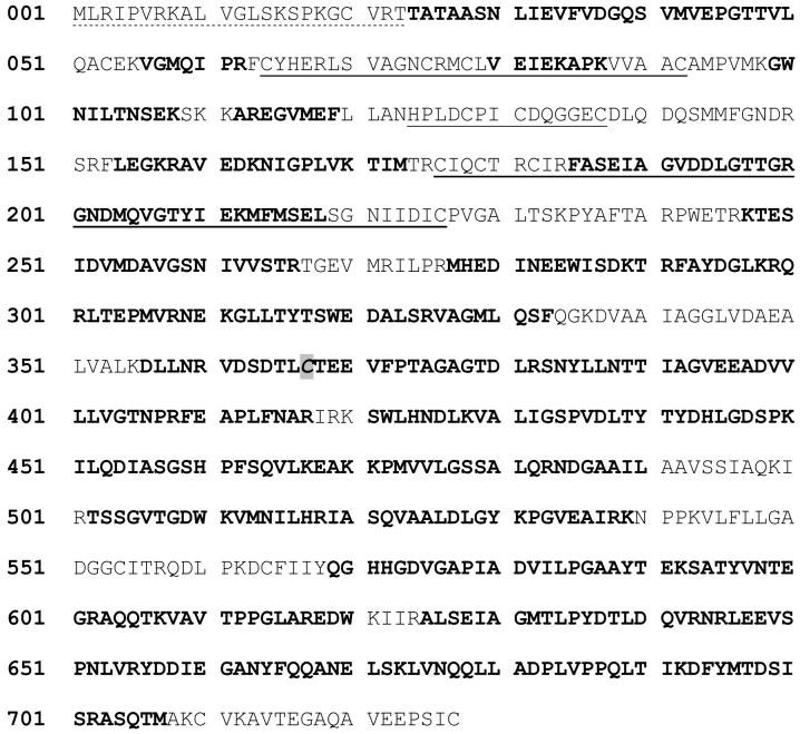
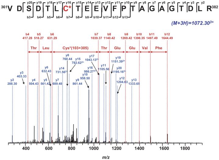
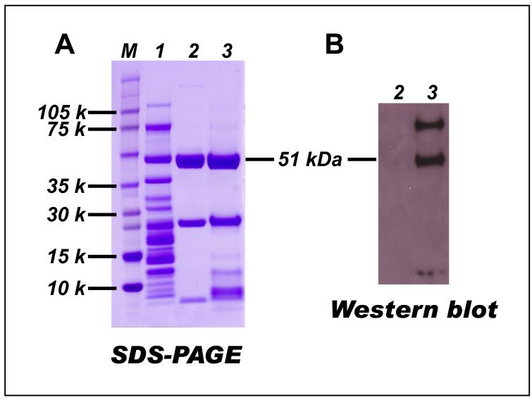
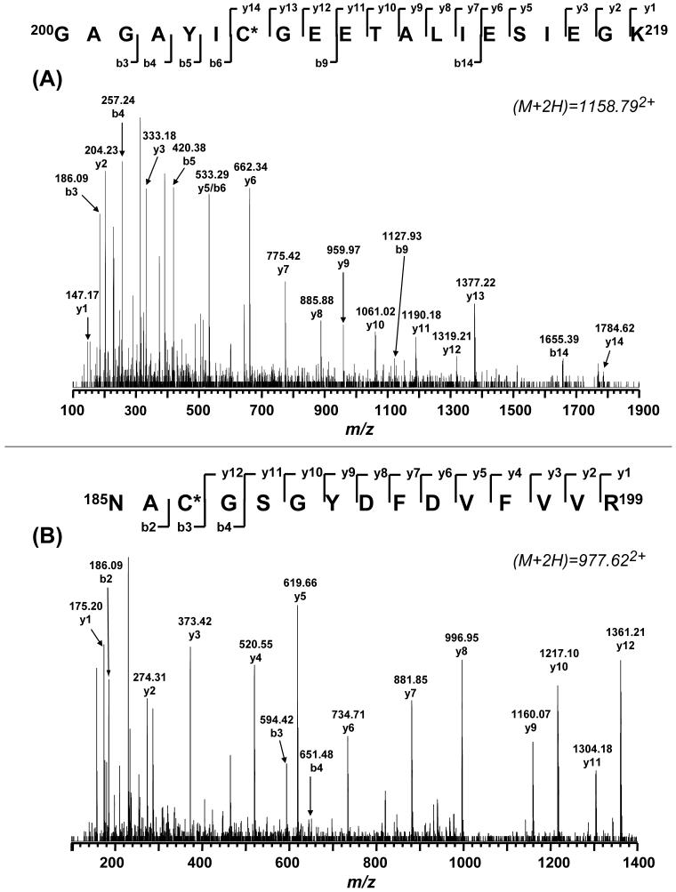
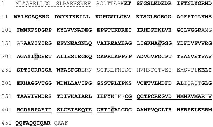
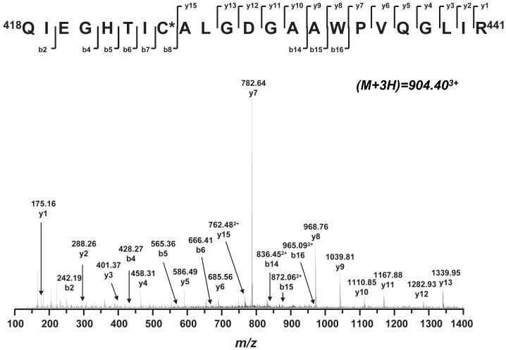
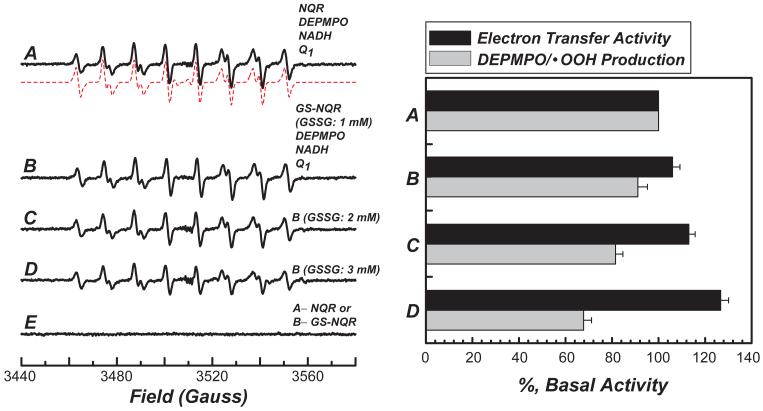
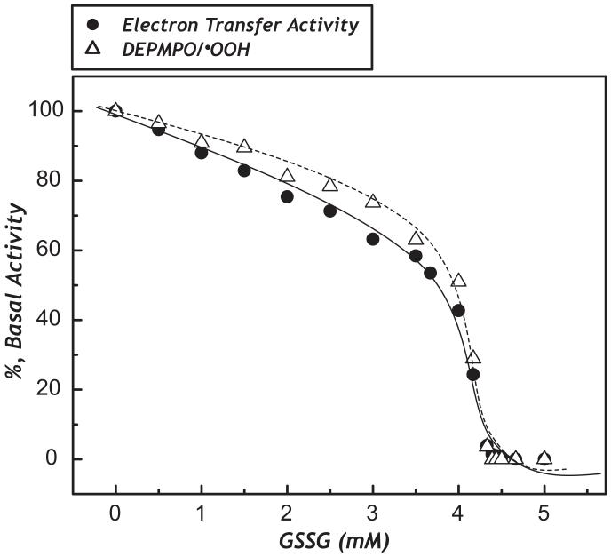
The generation of reactive oxygen species in mitochondria acts as a redox signal in triggering cellular events such as apoptosis, proliferation, and senescence. Overproduction of superoxide (O2*-) and O2*--derived oxidants changes the redox status of the mitochondrial GSH pool. An electron transport protein, mitochondrial complex I, is the major host of reactive/regulatory protein thiols. An important response of protein thiols to oxidative stress is to reversibly form protein mixed disulfide via S-glutathiolation. Exposure of complex I to oxidized GSH, GSSG, resulted in specific S-glutathiolation at the 51 kDa and 75 kDa subunits (Beer et al. (2004) J. Biol. Chem. 279, 47939-47951). Here, to investigate the molecular mechanism of S-glutathiolation of complex I, we prepared isolated bovine complex I under nonreducing conditions and employed the techniques of mass spectrometry and EPR spin trapping for analysis. LC/MS/MS analysis of tryptic digests of the 51 kDa and 75 kDa polypeptides from glutathiolated complex I (GS-NQR) revealed that two specific cysteines (C206 and C187) of the 51 kDa subunit and one specific cysteine (C367) of the 75 kDa subunit were involved in redox modifications with GS binding. The electron transfer activity (ETA) of GS-NQR in catalyzing NADH oxidation by Q1 was significantly enhanced. However, O2*- generation activity (SGA) mediated by GS-NQR suffered a mild loss as measured by EPR spin trapping, suggesting the protective role of S-glutathiolation in the intact complex I. Exposure of NADH dehydrogenase (NDH), the flavin subcomplex of complex I, to GSSG resulted in specific S-glutathiolation on the 51 kDa subunit. Both ETA and SGA of S-glutathiolated NDH (GS-NDH) decreased in parallel as the dosage of GSSG increased. LC/MS/MS analysis of a tryptic digest of the 51 kDa subunit from GS-NDH revealed that C206, C187, and C425 were glutathiolated. C425 of the 51 kDa subunit is a ligand residue of the 4Fe-4S N3 center, suggesting that destruction of 4Fe-4S is the major mechanism involved in the inhibition of NDH. The result also implies that S-glutathiolation of the 75 kDa subunit may play a role in protecting the 4Fe-4S cluster of the 51 kDa subunit from redox modification when complex I is exposed to redox change in the GSH pool.


Figures

Fig. 1
Fig. 1. Immunoblotting of the protein GSH mixed disulfide (PrSSG) of NQR (A) and NDH (B) using an anti-GSH monoclonal antibody
A, The NQR-derived PrSSG was induced by a thiol disulfide exchange reaction (Equation 1) from NQR (2.8 mg/ml) in PBS with various amounts (0-3 mM) of GSSG at room temperature for 1 h. B, NDH-derived PrSSG from the reaction (Equation 1) of NDH (0.23 mg/ml) in PBS with GSSG (1 mM) at room temperature for 1 h.
Fig. 2
Fig. 2. Amino acid sequence of the 75 kDa subunit precursor of NQR
The region labeled with bold represents the amino acid residues identified with LC/MS/MS. The underlined regions are the proposed sequence motif of 4Fe-4S binding (aa residues 64-92, 124-137, 176-226). The residues highlighted with gray color are involved in GS-binding (C367). The region labeled with a dotted underline is the signal peptide (residues 1-23), which acts as an import sequence and does not exist in the mature protein.
Fig. 3
Fig. 3. Tandem mass spectrum (MS/MS) of the triply protonated molecular ion of the GS-binding peptide (361VDSDTLC367TEEVFPTAAGTDLR382) of the 75 kDa subunit from GS-NQR
The sequence-specific ions are labeled as y and b ions on the spectrum. The amino acid residues involved in GS binding are identified by asterisks.
Fig. 4
Fig. 4. Preparation of the crude NADH dehydrogenase (NDH) from GS-NQR
GS-NQR was prepared according to the procedure (using 3 mM GSSG here) described in the legend of Fig. 1. 100 % ethanol was added to a final concentration of 9% (v/v), and the mixture was incubated at 40 °C for 10 min. The sample was then chilled in an ice-salt water bath for 10 min and subjected to centrifugation at 25,000 rpm for 30 min. The supernatant containing crude NDH was collected and concentrated with Centricon 30. A, SDS-PAGE: lane 1, isolated NQR (32 μg); lane 2, isolated native NADH dehydrogenase (7.5 μg); lane3, crude NADH dehydrogenase (12 μg). M represents a molecular weight marker. B, Western blot using a monoclonal antibody against GSH: lane 2, isolated native NADH dehydrogenase; lane 3, crude NADH dehydrogenase obtained from GS-NQR.
Fig. 5
Fig. 5. Tandem mass spectra (MS/MS) of the doubly protonated molecular ions of the GS-binding peptides (A) 200GAGAYIC206GEETALIESIEGK219 and (B) 185NAC187GSGYDFDVFVVR199 of the 51 kDa subunit from GS-NDH
The sequence-specific ions are labeled as y and b ions on the spectra. The amino acid residues involved in GS-binding are identified by asterisks.
Fig. 6
Fig. 6. Amino acid sequence of the precursor of NADH dehydrogenase 51 kDa subunit
The regions labeled with bold represents the amino acid residues identified with LC/MS/MS. The underlined regions are the sequence motif of 4Fe-4S binding (aa 379-425). The residues involved in GS-binding are highlighted with gray. The region labeled with a dotted underline is the signal peptide (aa 1-20), which acts as an import sequence and does not exist in the mature protein.
Fig. 7
Fig. 7. Tandem mass spectrum (MS/MS) of the triply protonated molecular ion of the GS-binding peptide (418QIETHTIC425ALGDGAAWPVQGLIR441) of the 51 kDa subunit from the GS-NDH
The sequence-specific ions are labeled as y and b ions on the spectrum. The amino acid residues involved in GS-binding are identified by asterisks.
Fig. 8
Fig. 8. Effect of protein S-glutathiolation of NQR on NQR-mediated superoxide generation and its electron transfer activity
The isolated NQR (2.8 mg/ml) in PBS was incubated with various concentrations of GSSG (0-3 mM) at room temperature for 1 h. The mixture was then subjected to dialysis against PBS for 8 h with one change of buffer at 4 °C. The protein concentration of dialysate containing NQR or GS-NQR was determined by the Lowry method (38). An aliquot of NQR or GS-NQR (127 μg/ml final concentration) was added to a mixture containing NADH (0.5 mM), Q1 (0.2 mM), DEPMPO (20 mM), and DTPA (1 mM) prior to EPR measurement. The DEPMPO/·OOH in each spectrum was quantitated by double integration of the simulation spectrum (dashed line)(7, 23). For measuring the electron transfer activity of NQR or GSN-QR, an aliquot of dialysate was withdrawn and assayed as described in the “Materials and Methods.” A, EPR spectrum obtained from a complete system containing NQR, NADH, Q1 and DEPMPO. B, the same as A except that NQR was replaced with GS-NQR. The GS-NQR was prepared using 1 mM GSSG. C, the same as B except that GS-NQR was prepared using 2 mM GSSG. D, the same as B except that GS-NQR was prepared using 3 mM GSSG. E, the same as A, but NQR was omitted from the system .
Fig. 9
Fig. 9. Effect of protein S-glutathiolation of NDH on NDH-mediated electron transfer activity its superoxide generation activity (dashed line)
The isolated NDH (0.23 mg/ml) in PBS was incubated with various concentrations of GSSG (0-5 mM) at room temperature for 1 h. Excess GSSG was removed by passing the sample through a MicroBioSpin-6 column. An appropriate amount of NDH or GS-NDH enzyme solution was withdrawn and subjected to ETA measurement as described in “Materials and Methods.” For measuring the superoxide generation by NDH or GS-NDH, the experimental approach of EPR was the same as that described in the legend of Fig. 8, except that Q1 was omitted from the reaction mixture. The absolute basal activity of NDH-mediated superoxide generation activity is 28.1 nmol O O2·- production min-1 mg-1 using 4-Hydroxy-2,2,6,6-tetramethylpiperidinyloxy (TEMPOL) as a standard for spin quantitation. Each data point represents the average of enzymatic assay from two batches of NDH preparation.

References

    1. Hirst J, Carroll J, Fearnley IM, Shannon RJ, Walker JE. The nuclear encoded subunits of complex I from bovine heart mitochondria. Biochim Biophys Acta. 2003;1604:135–50. - PubMed
    1. Carroll J, Fearnley IM, Shannon RJ, Hirst J, Walker JE. Analysis of the subunit composition of complex I from bovine heart mitochondria. Mol Cell Proteomics. 2003;2:117–26. - PubMed
    1. Walker JE. The NADH:ubiquinone oxidoreductase (complex I) of respiratory chains. Q Rev Biophys. 1992;25:253–324. - PubMed
    1. Galkin A, Brandt U. Superoxide radical formation by pure complex I (NADH:ubiquinone oxidoreductase) from Yarrowia lipolytica. J Biol Chem. 2005;280:30129–35. - PubMed
    1. Kudin AP, Bimpong-Buta NY, Vielhaber S, Elger CE, Kunz WS. Characterization of superoxide-producing sites in isolated brain mitochondria. J Biol Chem. 2004;279:4127–35. - PubMed

Publication types