

Neurotoxic lesions of ventrolateral prefrontal cortex impair object-in-place scene memory
- PMID: 17445247
- PMCID: PMC1855623
- DOI: 10.1111/j.1460-9568.2007.05468.x
Neurotoxic lesions of ventrolateral prefrontal cortex impair object-in-place scene memory
Abstract
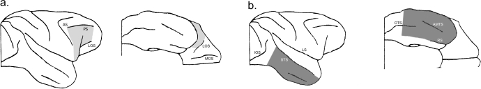
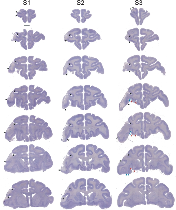
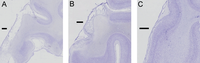
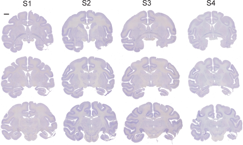
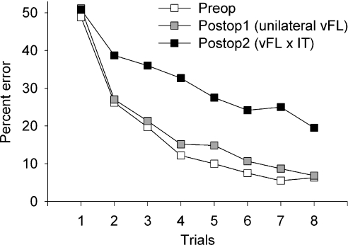
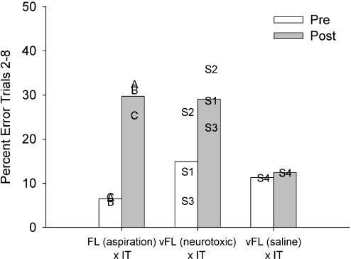
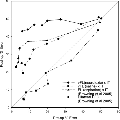
Disconnection of the frontal lobe from the inferotemporal cortex produces deficits in a number of cognitive tasks that require the application of memory-dependent rules to visual stimuli. The specific regions of frontal cortex that interact with the temporal lobe in performance of these tasks remain undefined. One capacity that is impaired by frontal-temporal disconnection is rapid learning of new object-in-place scene problems, in which visual discriminations between two small typographic characters are learned in the context of different visually complex scenes. In the present study, we examined whether neurotoxic lesions of ventrolateral prefrontal cortex in one hemisphere, combined with ablation of inferior temporal cortex in the contralateral hemisphere, would impair learning of new object-in-place scene problems. Male macaque monkeys learned 10 or 20 new object-in-place problems in each daily test session. Unilateral neurotoxic lesions of ventrolateral prefrontal cortex produced by multiple injections of a mixture of ibotenate and N-methyl-D-aspartate did not affect performance. However, when disconnection from inferotemporal cortex was completed by ablating this region contralateral to the neurotoxic prefrontal lesion, new learning was substantially impaired. Sham disconnection (injecting saline instead of neurotoxin contralateral to the inferotemporal lesion) did not affect performance. These findings support two conclusions: first, that the ventrolateral prefrontal cortex is a critical area within the frontal lobe for scene memory; and second, the effects of ablations of prefrontal cortex can be confidently attributed to the loss of cell bodies within the prefrontal cortex rather than to interruption of fibres of passage through the lesioned area.
Figures
Similar articles
-
Perseverative interference with object-in-place scene learning in rhesus monkeys with bilateral ablation of ventrolateral prefrontal cortex.Learn Mem. 2008 Feb 22;15(3):126-32. doi: 10.1101/lm.804508. Print 2008 Mar. Learn Mem. 2008. PMID: 18299439 Free PMC article.
-
Evidence for Mediodorsal Thalamus and Prefrontal Cortex Interactions during Cognition in Macaques.Cereb Cortex. 2015 Nov;25(11):4519-34. doi: 10.1093/cercor/bhv093. Epub 2015 May 15. Cereb Cortex. 2015. PMID: 25979086 Free PMC article.
-
Dissociable performance on scene learning and strategy implementation after lesions to magnocellular mediodorsal thalamic nucleus.J Neurosci. 2007 Oct 31;27(44):11888-95. doi: 10.1523/JNEUROSCI.1835-07.2007. J Neurosci. 2007. PMID: 17978029 Free PMC article.
-
Amnesia and neglect: beyond the Delay-Brion system and the Hebb synapse.Philos Trans R Soc Lond B Biol Sci. 1997 Oct 29;352(1360):1481-8. doi: 10.1098/rstb.1997.0135. Philos Trans R Soc Lond B Biol Sci. 1997. PMID: 9368937 Free PMC article. Review.
-
A neural system for human visual working memory.Proc Natl Acad Sci U S A. 1998 Feb 3;95(3):883-90. doi: 10.1073/pnas.95.3.883. Proc Natl Acad Sci U S A. 1998. PMID: 9448255 Free PMC article. Review.
Cited by
-
Behavioral control by the orbital prefrontal cortex: reversal of fortune.Nat Neurosci. 2013 Aug;16(8):984-5. doi: 10.1038/nn.3472. Nat Neurosci. 2013. PMID: 23887130 No abstract available.
-
Essential functions of primate frontopolar cortex in cognition.Proc Natl Acad Sci U S A. 2015 Mar 3;112(9):E1020-7. doi: 10.1073/pnas.1419649112. Epub 2015 Feb 17. Proc Natl Acad Sci U S A. 2015. PMID: 25691741 Free PMC article.
-
A conserved pattern of differential expansion of cortical areas in simian primates.J Neurosci. 2013 Sep 18;33(38):15120-5. doi: 10.1523/JNEUROSCI.2909-13.2013. J Neurosci. 2013. PMID: 24048842 Free PMC article.
-
The magnocellular mediodorsal thalamus is necessary for memory acquisition, but not retrieval.J Neurosci. 2008 Jan 2;28(1):258-63. doi: 10.1523/JNEUROSCI.4922-07.2008. J Neurosci. 2008. PMID: 18171943 Free PMC article.
-
Addition of fornix transection to frontal-temporal disconnection increases the impairment in object-in-place memory in macaque monkeys.Eur J Neurosci. 2008 Apr;27(7):1814-22. doi: 10.1111/j.1460-9568.2008.06140.x. Eur J Neurosci. 2008. PMID: 18380673 Free PMC article.
References
-
- Baxter MG, Murray EA. Reinterpreting the behavioural effects of amygdala lesions in nonhuman primates. In: Aggleton JP, editor. The Amygdala: a Functional Analysis. Oxford, UK: Oxford University Press; 2000. pp. 545–568.
-
- Browning PG, Easton A, Buckley MJ, Gaffan D. The role of prefrontal cortex in object-in-place learning in monkeys. EurJNeurosci. 2005;22:3281–3291. - PubMed
-
- Browning PG, Easton A, Gaffan D. Frontal-Temporal Disconnection Abolishes Object Discrimination Learning Set in Macaque Monkeys. CerebCortex. 2006 Published online May 17, 2006 [doi: 10.1093/cercor/bhk039] - PubMed
-
- Bussey TJ, Wise SP, Murray EA. Interaction of ventral and orbital prefrontal cortex with inferotemporal cortex in conditional visuomotor learning. BehavNeurosci. 2002;116:703–715. - PubMed
-
- Gaffan D. Scene-specific memory for objects: a model of episodic memory impairment in monkeys with fornix transection. JCognNeurosci. 1994;6:305–320. - PubMed
Publication types
MeSH terms
Grants and funding
LinkOut - more resources
Full Text Sources
Medical
