Comparative Study
. 2007 Jun 15;73(12):1873-86.
doi: 10.1016/j.bcp.2007.03.002. Epub 2007 Mar 12.

Multidrug-resistant cancer cells are preferential targets of the new antineoplastic lanthanum compound KP772 (FFC24)

Affiliations
Comparative Study

Multidrug-resistant cancer cells are preferential targets of the new antineoplastic lanthanum compound KP772 (FFC24)

P Heffeter et al. Biochem Pharmacol. .

Abstract

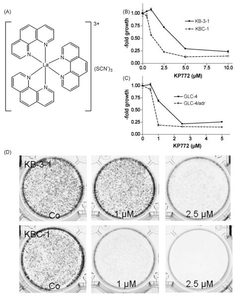
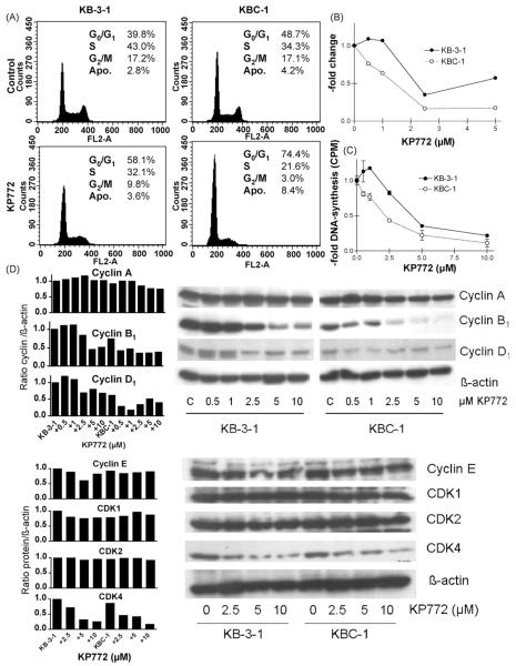
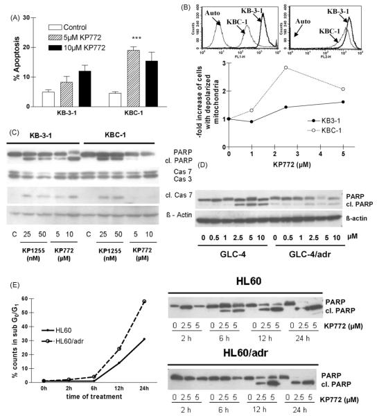
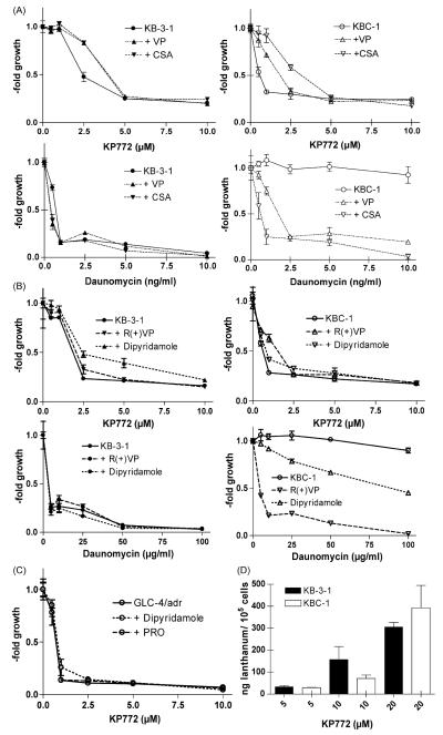
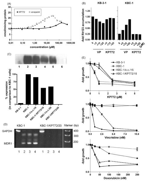
Recently, we have introduced [tris(1,10-phenanthroline)lanthanum(III)] trithiocyanate (KP772, FFC24) as a new lanthanum compound which has promising anticancer properties in vivo and in vitro. Aim of this study was to investigate the impact of ABC transporter-mediated multidrug resistance (MDR) on the anticancer activity of KP772. Here, we demonstrate that all MDR cell models investigated, overexpressing ABCB1 (P-glycoprotein), ABCC1 (multidrug resistance protein 1), or ABCG2 (breast cancer resistance protein) either due to drug selection or gene transfection, were significantly hypersensitive against KP772. Using ABCB1-overexpressing KBC-1 cells as MDR model, KP772 hypersensitivity was demonstrated to be based on stronger apoptosis induction and/or cell cycle arrest at unaltered cellular drug accumulation. KP772 did neither stimulate ABCB1 ATPase activity nor alter rhodamine 123 accumulation arguing against a direct interaction with ABCB1. Accordingly, several drug resistance modulators did not sensitize but rather protect MDR cells against KP772-induced cytotoxicity. Moreover, long-term KP772 treatment of KBC-1 cells at subtoxic concentrations led within 20 passages to a complete loss of drug resistance based on blocked MDR1 gene expression. When exposing parental KB-3-1 cells to subtoxic, stepwise increasing KP772 concentrations, we observed, in contrast to several other metallo-drugs, no acquisition of KP772 resistance. Summarizing, our data demonstrate that KP772 is hyperactive in MDR cells and might have chemosensitizing properties by blocking ABCB1 expression. Together with the disability of tumor cells to acquire KP772 resistance, our data suggest that KP772 should be especially active against notoriously drug-resistant tumor types and as second line treatment after standard chemotherapy failure.


Figures

Fig. 1
Fig. 1
(A) [Tris(1,10-phenanthroline)lanthanum(III)] trithiocyanate (KP772; FFC24). KP772-induced cytotoxicity against (B) KB-3-1 or (C) GLC-4 and their chemoresistant sublines KBC-1 (P-gp-overexpressing) or GLC-4/adr (MRP1-overexpressing) was measured using MTT assay after 72 h drug exposure. (D) Clonogenic survival of KB-3-1 and KBC-1 cells was determined after exposure to the indicated concentrations of KP772 for 6 days. Cell colonies were visualised by crystal violet staining.
Fig. 2
Fig. 2
Impact of KP772 on cell cycle progression and DNA synthesis of KB-3-1 and ABCB1-overexpressing KBC-1 cells. (A) Changes in the cell cycle distribution of the indicated cell lines treated with 5 μM KP772 for 24 h were analysed by PI staining and FACS. Percentages of cells in G0/G1, S and G2/M phases of the cell cycle as well as apoptotic cells (Apo) are indicated. (B) Changes in the proportion of cells in G2/M phase of the cell cycle at increasing doses of KP772 are shown. The amount of G2/M cells in the untreated control group was set as 1. One of three experiments delivering comparable results is shown. (C) DNA synthesis was determined by 3H-thymidine incorporation after 24 h treatment with KP772 at the indicated concentrations. Values given are means ± S.D. from at least two independent experiments performed in triplicates. (D) The impact of the indicated drug concentrations on the expression pattern of cyclin A, B1, D1, E and CDK1, 2, 4 after a 24 h treatment was analysed by Western blot (right) followed by densiometric evaluation (left). β-Actin was used as loading control. Antibodies are described under Section 2.
Fig. 3
Fig. 3
Induction of apoptosis in KB-3-1 and ABCB1-overexpressing KBC-1 cells after treatment with KP772 for 24 h. (A) Percentages of apoptotic nuclei in untreated controls and cells treated with 5 and 10 μM KP772 were determined microscopically after DAPI staining. Three hundred to 500 nuclei of at least two cytospin slides for each concentration and cell line were counted. (B) Accumulation of green-fluorescent cytoplasmic (FL-1, left) and red-fluorescent mitochondrial (FL-2, right) JC-1 in the indicated cell lines is shown in the upper panel. The lower panel indicates loss of mitochondrial membrane potential after treatment with KP772. The proportions of apoptotic KB-3-1 and KBC-1 cells after treatment with the indicated drug concentrations are shown normalised to the untreated control. (C) Caspase-induced cleavage of PARP, caspase 7 and caspase 3 in KB-3-1 and KBC-1 cells after treatment with KP772 at the indicated concentrations was determined via Western blotting. The bismuth compound KP1255 was used as positive control. Antibodies used are described under Section 2. (D) Caspase-induced cleavage of PARP in GLC-4 and ABCC1- and LRP-overexpressing GLC-4/adr cells after treatment with KP772 at the indicated concentrations was determined via Western blotting. (E) After treatment with KP772 for the indicated time periods, HL60 and ABCC1-overexpressing HL60/adr cells were analysed for apoptosis induction by both Western blotting and FACS. Immunoblots (right panel) show the time-dependent cleavage of PARP by treatment with 2.5 and 5 μM KP772. For FACS analysis cells treated with 2.5 μM KP772 were fixed and stained with ethanol and PI, respectively. Percentages of the apoptotic subG0/G1 compartment were calculated by Cell Quest Software.
Fig. 4
Fig. 4
Impact of MDR modulators on KP772 anticancer activity. KB-3-1 cells and the ABCB1-overexpressing subline KBC-1 were incubated for 72 h with increasing concentrations of KP772 in combination with the ABCB1/ABCC modulators (A) VP (10 μM) and CSA (1 μM) and (B) R(+)VP (10 μM) and dipyridamole (10 μM) as indicated. Values given are means ± S.D. of one representative experiment performed in triplicate. At least three experiments were done delivering comparable results. (C) Impact of dipyridamole (10 μM) and PRO (1 mM) on KP772-treated GLC-4/adr cells. (D) Lanthanum levels of KB-3-1 (full bars) and KBC-1 cells (open bars) were measured by ICP-MS after 1 h incubation with the indicated KP772 concentrations. Means ± S.D. of at least three experiments are given.
Fig. 5
Fig. 5
Impacts of KP772 on ABCB1 expression and function. (A) The impact on ABCB1 ATPase activity was determined by analysing the rate of ATP hydrolysis in ABCB1-containing plasma membrane vesicles [9] at increasing concentrations of KP772 (straight line) or VP (dashed line). The concentration–response curves were fitted to the data points by non-linear regression analysis. (B) Rh123 accumulation in KB-3-1 and KBC-1 cells with and without coadministration of KP772 and VP at the indicated concentrations was measured after 1 h drug exposure by FACS analysis. Data were normalised to Rh123 accumulation of untreated KB-3-1 cells. One of three experiments delivering comparable results is shown. (C) ABCB1 expression levels of KB-3-1 (lane 1), KBC-1 (lane 2), and KBC-1 cells cultured without colchicine selection for 10 passages (KBC-1/o.c./10; lane 3), and 20 passages (KBC-1/o.c./20; lane 5) or under exposure to 0.7 μM KP772 for the identical time periods (KBC-1/KP772/10; lane 4 and KBC-1/KP772/20; lane 6, respectively) were measured in membrane-enriched fractions by Western blot and quantified by Molecular Analyst software (Biorad). (D) ABCB1 mRNA expression in KBC-1 and KBC-1/772/20 cells was analysed by RT-PCR. Amplification products obtained using GAPDH-specific (358 bp product; 25 cycles, lane 1) and ABCB1-specific (167 bp product; 25, 30, 35 PCR cycles, lanes 2–4) oligonucleotide primers, respectively, were separated by acrylamide gel electrophoresis and stained by ethidium bromide. (E) Concentration–response curves were established for the indicated drugs in KB-3-1, KBC-1, KBC-1/o.c./15 and KBC-1/KP772/15 cells. Following 72 h drug exposure, cell viability was determined by MTT assays.
Fig. 6
Fig. 6
Morphologic changes in KB-3-1 cells after 8-month selection against KP772. Drug concentrations were stepwise, slowly increased (compare Section 2) until the indicated levels were reached. Photomicrographs shown were taken by using a 10× objective and phase contrast settings using Nikon Eclipse TE300 (Nikon Instruments, Japan). Bar: 50 μm.

References

    1. Dean M, Fojo T, Bates S. Tumour stem cells and drug resistance. Nat Rev Cancer. 2005;5:275–84. - PubMed
    1. Szakacs G, Paterson JK, Ludwig JA, Booth-Genthe C, Gottesman MM. Targeting multidrug resistance in cancer. Nat Rev Drug Discov. 2006;5:219–34. - PubMed
    1. Heffeter P, Jakupec MA, Korner W, Wild S, von Keyserlingk NG, Elbling L, et al. Anticancer activity of the lanthanum compound [tris(1,10-phenanthroline)lanthanum(III)]trithiocyanate (KP772; FFC24) Biochem Pharmacol. 2006;71:426–40. - PubMed
    1. Shen DW, Cardarelli C, Hwang J, Cornwell M, Richert N, Ishii S, et al. Multiple drug-resistant human KB carcinoma cells independently selected for high-level resistance to colchicine, adriamycin, or vinblastine show changes in expression of specific proteins. J Biol Chem. 1986;261:7762–70. - PubMed
    1. McGrath T, Center MS. Mechanisms of multidrug resistance in HL60 cells: evidence that a surface membrane protein distinct from P-glycoprotein contributes to reduced cellular accumulation of drug. Cancer Res. 1988;48:3959–63. - PubMed

Publication types

MeSH terms

Substances