

Neurocognitive and neuroimaging correlates of pediatric traumatic brain injury: a diffusion tensor imaging (DTI) study
- PMID: 17446039
- PMCID: PMC2887608
- DOI: 10.1016/j.acn.2007.03.004
Neurocognitive and neuroimaging correlates of pediatric traumatic brain injury: a diffusion tensor imaging (DTI) study
Abstract
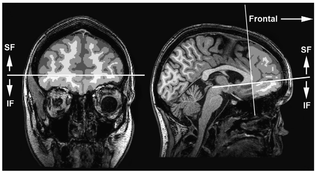
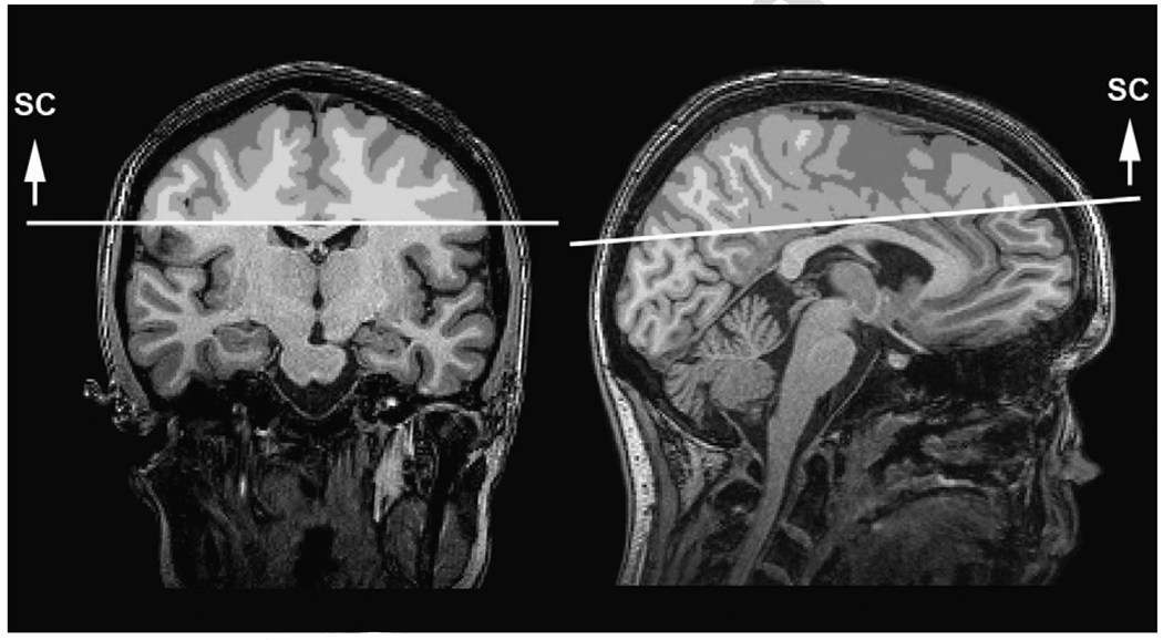
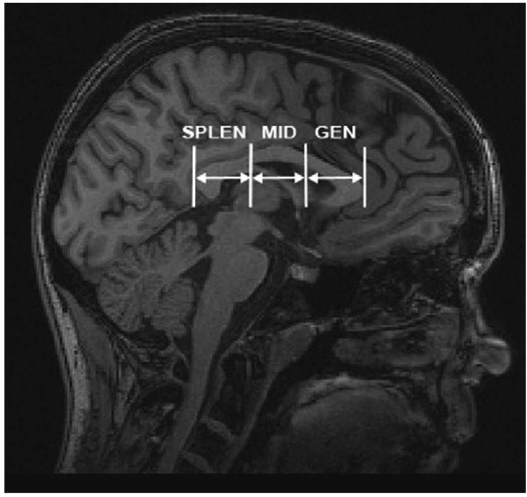
This study examined the sensitivity of diffusion tensor imaging (DTI) to microstructural white matter (WM) damage in mild and moderate pediatric traumatic brain injury (TBI). Fourteen children with TBI and 14 controls ages 10-18 had DTI scans and neurocognitive evaluations at 6-12 months post-injury. Groups did not differ in intelligence, but children with TBI showed slower processing speed, working memory and executive deficits, and greater behavioral dysregulation. The TBI group had lower fractional anisotropy (FA) in three WM regions: inferior frontal, superior frontal, and supracallosal. There were no group differences in corpus callosum. FA in the frontal and supracallosal regions was correlated with executive functioning. Supracallosal FA was also correlated with motor speed. Behavior ratings showed correlations with supracallosal FA. Parent-reported executive deficits were inversely correlated with FA. Results suggest that DTI measures are sensitive to long-term WM changes and associated with cognitive functioning following pediatric TBI.
Figures
Similar articles
-
Diffusion tensor imaging characteristics of the corpus callosum in mild, moderate, and severe traumatic brain injury.AJNR Am J Neuroradiol. 2008 Oct;29(9):1730-5. doi: 10.3174/ajnr.A1213. Epub 2008 Jul 10. AJNR Am J Neuroradiol. 2008. PMID: 18617586 Free PMC article.
-
Working memory and corpus callosum microstructural integrity after pediatric traumatic brain injury: a diffusion tensor tractography study.J Neurotrauma. 2013 Oct 1;30(19):1609-19. doi: 10.1089/neu.2013.2934. Epub 2013 Aug 24. J Neurotrauma. 2013. PMID: 23627735 Free PMC article.
-
Longitudinal changes in the corpus callosum following pediatric traumatic brain injury.Dev Neurosci. 2010;32(5-6):361-73. doi: 10.1159/000317058. Epub 2010 Oct 14. Dev Neurosci. 2010. PMID: 20948181 Free PMC article.
-
The relationship between diffusion tensor imaging findings and cognitive outcomes following adult traumatic brain injury: A meta-analysis.Neurosci Biobehav Rev. 2018 Sep;92:93-103. doi: 10.1016/j.neubiorev.2018.05.023. Epub 2018 May 24. Neurosci Biobehav Rev. 2018. PMID: 29803527 Review.
-
Neuroimaging in pediatric traumatic brain injury: current and future predictors of functional outcome.Dev Disabil Res Rev. 2009;15(2):117-23. doi: 10.1002/ddrr.62. Dev Disabil Res Rev. 2009. PMID: 19489082 Free PMC article. Review.
Cited by
-
Early microstructural and metabolic changes following controlled cortical impact injury in rat: a magnetic resonance imaging and spectroscopy study.J Neurotrauma. 2011 Oct;28(10):2091-102. doi: 10.1089/neu.2010.1739. Epub 2011 Sep 29. J Neurotrauma. 2011. PMID: 21761962 Free PMC article.
-
Assessment of White Matter Integrity after Pediatric Traumatic Brain Injury.J Neurotrauma. 2020 Oct 15;37(20):2188-2197. doi: 10.1089/neu.2019.6691. Epub 2020 May 15. J Neurotrauma. 2020. PMID: 32253971 Free PMC article.
-
Moderate injury in motor-sensory cortex causes behavioral deficits accompanied by electrophysiological changes in mice adulthood.PLoS One. 2017 Feb 14;12(2):e0171976. doi: 10.1371/journal.pone.0171976. eCollection 2017. PLoS One. 2017. PMID: 28196142 Free PMC article.
-
White matter and reading deficits after pediatric traumatic brain injury: A diffusion tensor imaging study.Neuroimage Clin. 2015 Oct 19;9:668-77. doi: 10.1016/j.nicl.2015.10.009. eCollection 2015. Neuroimage Clin. 2015. PMID: 26740920 Free PMC article.
-
The young brain and concussion: imaging as a biomarker for diagnosis and prognosis.Neurosci Biobehav Rev. 2012 Jul;36(6):1510-31. doi: 10.1016/j.neubiorev.2012.03.007. Epub 2012 Mar 28. Neurosci Biobehav Rev. 2012. PMID: 22476089 Free PMC article. Review.
References
-
- Anderson V, Catroppa C, Morse S, Haritou F, Rosenfeld J. Attentional and processing skills following traumatic brain injury in early childhood. Brain Injury. 2005;19(9):699–710. - PubMed
-
- Anderson V, Fenwick T, Manly T, Robertson I. Attentional skills following traumatic brain injury in childhood: A componential analysis. Brain Injury. 1998;12(11):937–949. - PubMed
-
- Anderson V, Morse SA, Catroppa C, Haritou F, Rosenfeld JV. Thirty month outcome from early childhood head injury: A prospective analysis of neurobehavioral recovery. Brain. 2004;127(Pt 12):2608–2620. - PubMed
-
- Asarnow RF, Satz P, Light R, Lewis R, Neumann E. Behavior problems and adaptive functioning in children with mild and severe closed head injury. Journal of Pediatric Psychology. 1991;16(5):543–555. - PubMed
Publication types
MeSH terms
Grants and funding
LinkOut - more resources
Full Text Sources
Medical
