

Feedforward versus feedback modulation of human vestibular-evoked balance responses by visual self-motion information
- PMID: 17446222
- PMCID: PMC2075304
- DOI: 10.1113/jphysiol.2007.132092
Feedforward versus feedback modulation of human vestibular-evoked balance responses by visual self-motion information
Abstract
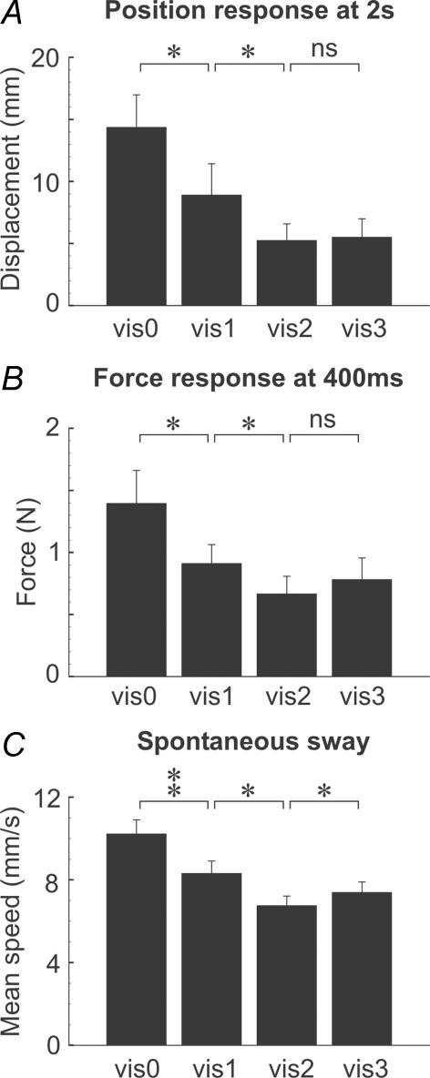
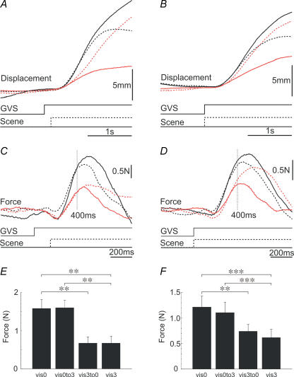
Visual information modulates the balance response evoked by a pure vestibular perturbation (galvanic vestibular stimulation, GVS). Here we investigate two competing hypotheses underlying this visual-vestibular interaction. One hypothesis assumes vision acts in a feedforward manner by altering the weight of the vestibular channel of balance control. The other assumes vision acts in a feedback manner through shifts in the retinal image produced by the primary response. In the first experiment we demonstrate a phenomenon that is predicted by both hypotheses: the GVS-evoked balance response becomes progressively smaller as the amount of visual self-motion information is increased. In the second experiment we independently vary the pre-stimulus and post-stimulus visual environments. The rationale is that feedback effects would depend only upon the post-stimulus visual environment. Although the post-stimulus visual environment did affect later parts of the response (after approximately 400 ms), the pre-stimulus visual environment had a strong influence on the size of the early part of the response. We conclude that both feedforward and feedback mechanisms act in concert to modulate the GVS-evoked response. We suggest this dual interaction that we observe between visual and vestibular channels is likely to apply to all sensory channels that contribute to balance control.
Figures


References
-
- Brandt T, Bartenstein P, Janek A, Dieterich M. Reciprocal inhibitory visual–vestibular interaction. Visual motion stimulation deactivates the parieto-insular vestibular cortex. Brain. 1998;121:1749–1758. - PubMed
-
- Breson K, Elberling C, Fangel J. Galvanic nystagmography. Acta Otolaryngol. 1971;71:449–455. - PubMed
-
- Britton TC, Day BL, Brown P, Rothwell JC, Thompson PD, Marsden CD. Postural electromyographic responses in the arm and leg following galvanic vestibular stimulation in man. Exp Brain Res. 1993;94:143–151. - PubMed
-
- Bronstein AM, Buckwell D. Automatic control of postural sway by visual motion parallax. Exp Brain Res. 1997;113:243–248. - PubMed
-
- Cenciarini M, Peterka RJ. Stimulus-dependent changes in the vestibular contribution to human postural control. J Neurophysiol. 2006;95:2733–2750. - PubMed
Publication types
MeSH terms
Grants and funding
LinkOut - more resources
Full Text Sources