

Neurovascular invasion at the osteochondral junction and in osteophytes in osteoarthritis
- PMID: 17446239
- PMCID: PMC2111605
- DOI: 10.1136/ard.2006.063354
Neurovascular invasion at the osteochondral junction and in osteophytes in osteoarthritis
Abstract
Background: Normal adult articular cartilage is thought to be avascular and aneural.
Objective: To describe neurovascular structures at the osteochondral junction and in osteophytes in tibiofemoral osteoarthritis (OA) displaying a range of severity of cartilage changes.
Methods: Articular surfaces were obtained from 40 patients at total knee joint replacement surgery for tibiofemoral OA (TKR) and seven patients post mortem (PM). Antibodies directed against CD34 (vascular endothelium), protein gene product 9.5 (pan-neuronal marker), substance P and calcitonin gene-related peptide (sensory nerves) and C-flanking peptide of neuropeptide Y (sympathetic nerves) were used to localise blood vessels and nerves by immunohistochemistry. Severity of OA cartilage changes was graded histologically.
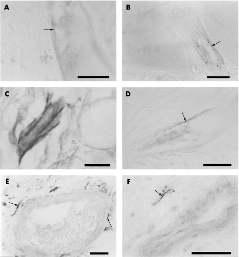
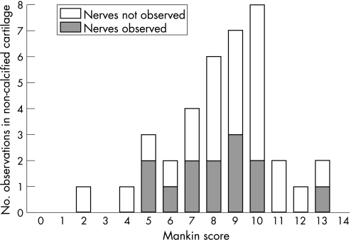
Results: TKR and PM samples displayed a range of OA cartilage changes including tidemark breaching by vascular channels. Sympathetic and sensory nerves were both present within vascular channels in the articular cartilage, in both mild and severe OA. Perivascular and free nerve fibres, and nerve trunks were observed within the subchondral bone marrow and within the marrow cavities of osteophytes. Sensory and sympathetic nerves displayed similar distributions in each region studied.
Conclusion: Vascularisation and the associated innervation of articular cartilage may contribute to tibiofemoral pain in OA across a wide range of structural disease severity.
Conflict of interest statement
Competing interests: None.

References
-
- Doherty M. Pathogenesis. Introduction: the concept of osteoarthritis as failure of the diarthrodial joint. In: Brandt KD, Doherty M, Lohmander S, eds. Osteoarthritis. 1st edn. Oxford: Oxford University Press 199870–74.
-
- Duncan H, Jundt J, Riddle J M, Pitchford W, Christopherson T. The tibial subchondral plate. A scanning electron microscopic study. J Bone Joint Surg [Am] 1987691212–1220. - PubMed
-
- Hashimoto S, Creighton‐Achermann L, Takahashi K, Amiel D, Coutts R D, Lotz M. Development and regulation of osteophyte formation during experimental osteoarthritis. Osteoarthritis Cartilage 200210180–187. - PubMed
-
- Gronblad M, Liesi P, Korkala O, Karaharju E, Polak J. Innervation of human bone periosteum by peptidergic nerves. Anat Rec 1984209297–299. - PubMed
Publication types
MeSH terms
Substances
LinkOut - more resources
Full Text Sources
Other Literature Sources
Miscellaneous
