. 2007 Aug 15;110(4):1147-52.
doi: 10.1182/blood-2007-02-077099. Epub 2007 Apr 19.

The spectrum of mutations in the PCFT gene, coding for an intestinal folate transporter, that are the basis for hereditary folate malabsorption

Affiliations

The spectrum of mutations in the PCFT gene, coding for an intestinal folate transporter, that are the basis for hereditary folate malabsorption

Rongbao Zhao et al. Blood. .

Abstract

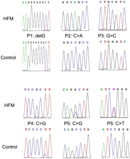
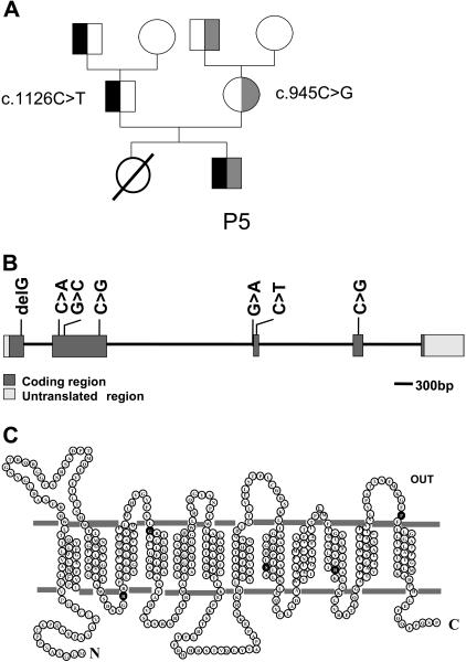
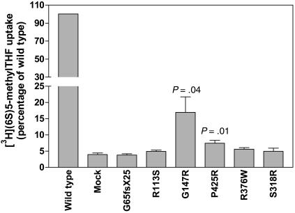
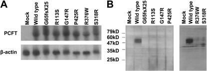
Hereditary folate malabsorption (HFM) is a rare autosomal recessive disorder caused by impaired intestinal folate absorption and impaired folate transport into the central nervous system. Recent studies in 1 family revealed that the molecular basis for this disorder is a loss-of-function mutation in the PCFT gene encoding a proton-coupled folate transporter. The current study broadens the understanding of the spectrum of alterations in the PCFT gene associated with HFM in 5 additional patients. There was no racial, ethnic, or sex pattern. A total of 4 different homozygous mutations were detected in 4 patients; 2 heterozygous mutations were identified in the fifth patient. Mutations involved 4 of the 5 exons, all at highly conserved amino acid residues. A total of 4 of the mutated transporters resulted in a complete loss of transport function, primarily due to decreased protein stability and/or defects in membrane trafficking, while 2 of the mutated carriers manifested residual function. Folate transport at low pH was markedly impaired in transformed lymphocytes from 2 patients. These findings further substantiate the role that mutations in PCFT play in the pathogenesis of HFM and will make possible rapid diagnosis and treatment of this disorder in infants, and prenatal diagnosis in families that carry a mutated gene.


Figures

Figure 1
Figure 1
Representative chromatograms of DNA sequence data. Top panels (HFM) represent the mutated DNA in patients with HFM, whereas the bottom panels (control) are corresponding wild-type sequences.
Figure 2
Figure 2
Pedigree of a family with HFM; localization of PCFT mutations. (A) Pedigree of the family of patient P5. The black color indicates the c.1126C > T mutation, and the gray color indicates the c.954C > G mutation. (B) The genomic organization of the PCFT gene and the location of mutations detected in patients with HFM. (C) The location of amino acid substitutions (●) associated with mutations in PCFT gene in a topological structure predicted by online databases (ExPASy Proteomics Tools and Transport Classification Database).
Figure 3
Figure 3
[3H]5-methylTHF uptake in HeLa cells transiently transfected with the cDNA of PCFT mutants. Uptake of 0.5 μM [3H](6S)5-methylTHF was assessed at pH 5.5 and 37°C over 2 minutes; P values reflect differences in activities of the mutated carriers as compared with the mock transfectants. The data are the mean plus or minus SEM from 4 independent experiments.
Figure 4
Figure 4
Northern and Western blot analyses of PCFT mutants. (A) Northern blot analysis. Total RNA (20 μg) was fractionated on a formaldehyde-agarose gel, and the blot was successively probed with a PCFT fragment and β-actin. The images represent 1 of 2 independent experiments. (B) Western blot analysis. An equal amount of lysate (20 μg) was loaded on SDS gels, and the blots were probed with a peptide antibody directed to the C-terminus of PCFT. The numbers on the left indicate the molecular sizes of the protein ladder. The left and right panels represent 2 different analyzes each with mock and wild-type PCFT as controls. Each blot is representative of 3 separate experiments.
Figure 5
Figure 5
Immunofluorescence staining of PCFT. HeLa cells were transiently transfected with cDNA of wild-type and PCFT mutants and labeled with a rabbit antibody targeted to human PCFT. (A) Wild-type PCFT. (B) P425R. (C) R376W. (D) S318R. Green fluorescence indicates localization of PCFT, while the blue fluorescence (panels A-B) or red fluorescence (panels C,D) shows nucleus counterstained by DAPI or PI, respectively. The image is a representative view from at least 3 experiments. The same antibody was used for immunohistochemical and Western blot (Figure 4B) analyses. The image was obtained on Olympus 1X70 inverted Epifluorescence Microscope (Olympus) with a 60×/1.40 oil objective, recorded on a Sensicam QE cooled CCD Camera (Applied Scientific Instrumentation), acquired with software IP Lab 4.0 (BD Biosciences) and further processed with Photoshop CS (Adobe Systems).
Figure 6
Figure 6
[3H]5-methylTHF uptake at low pH in lymphocytes derived from patients P6 and P7 and their parents. Uptake of 0.5 μM [3H](6S)5-methylTHF at pH 5.5 was assessed over 2 minutes. F+/− and M+/− indicate cells derived from father and mother, respectively, who each carry 1 mutated PCFT allele. The data are the mean plus or minus SEM from 3 independent experiments.

References

    1. Geller J, Kronn D, Jayabose S, Sandoval C. Hereditary folate malabsorption: family report and review of the literature. Medicine (Baltimore) 2002;81:51–68. - PubMed
    1. Poncz M, Cohen A. Long-term treatment of congenital folate malabsorption. J Pediatr. 1996;129:948. - PubMed
    1. Corbeel L, Van den Berghe G, Jaeken J, Van Tornout J, Eeckels R. Congenital folate malabsorption. Eur J Pediatr. 1985;143:284–290. - PubMed
    1. Jebnoun S, Kacem S, Mokrani CH, Chabchoub A, Khrouf N, Zittoun J. A family study of congenital malabsorption of folate. J Inherit Metab Dis. 2001;24:749–750. - PubMed
    1. Qiu A, Jansen M, Sakaris A, et al. Identification of an intestinal folate transporter and the molecular basis for hereditary folate malabsorption. Cell. 2006;127:917–928. - PubMed

Publication types

MeSH terms