. 2007 May 2;26(9):2327-38.
doi: 10.1038/sj.emboj.7601679. Epub 2007 Apr 19.

Translation of nonSTOP mRNA is repressed post-initiation in mammalian cells

Affiliations

Translation of nonSTOP mRNA is repressed post-initiation in mammalian cells

Nobuyoshi Akimitsu et al. EMBO J. .

Abstract

We investigated the fate of aberrant mRNAs lacking in-frame termination codons (called nonSTOP mRNA) in mammalian cells. We found that translation of nonSTOP mRNA was considerably repressed although a corresponding reduction of mRNA was not observed. The repression appears to be post-initiation since (i) repressed nonSTOP mRNAs were associated with polysomes, (ii) translation of IRES-initiated and uncapped nonSTOP mRNA were repressed, and (iii) protein production from nonSTOP mRNA associating with polysomes was significantly reduced when used to program an in vitro run-off translation assay. NonSTOP mRNAs distributed into lighter polysome fractions compared to control mRNAs encoding a stop codon, and a significant amount of heterogeneous polypeptides were produced during in vitro translation of nonSTOP RNAs, suggesting premature termination of ribosomes translating nonSTOP mRNA. Moreover, a run-off translation assay using hippuristanol and RNAse protection assays suggested the presence of a ribosome stalled at the 3' end of nonSTOP mRNAs. Taken together, these data indicate that ribosome stalling at the 3' end of nonSTOP mRNAs can block translation by preventing upstream translation events.


Figures

Figure 1
Figure 1
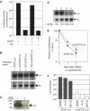
Expression of nonSTOP GFP in HeLa cells. (A) Schematic drawing of GFP reporter genes. Right-angled arrows indicate the CMV promoter. Open boxes indicate the coding frame and shaded boxes indicate the polyadenylation signal. The presence of the GFP termination codon is indicated by a STOP sign. The lengths of the ORFs are indicated. (B) Fluorescence microscopic observation of cells transfected with the indicated pEGFP-N1-derived plasmid and pDsRed2 plasmid. Upper panels are results of observation with the Green fluorescence filter set (U-MGFP, Olympus). Lower panels are results of observation with the red fluorescence filter set (U-MWIG, Olympus). (C) Northern blot analysis of total RNA extracted from HeLa cells transfected with either pGFP STOP (lane 1 and 3) or pGFP nonSTOP (lanes 2 and 4) plasmid. In all, 1 μg reporter plasmid (lanes 1 and 2) or 100 ng plasmid (lanes 3 and 4) was transfected into cells. Total RNA (10 μg) was resolved on a 1.2% agarose–formaldehyde gel and probed with 32P-labeled GFP (upper panel) or neomycin phosphotransferase (Neo) (lower panel). The relative expression levels of GFP mRNA normalized to Neo mRNA are indicated below the panels. (D) Western blot hybridization analysis of extracts prepared from HeLa cells transfected with either the pGFP STOP (lane 1) or pGFP nonSTOP (lane 2) plasmid. Extracts were resolved by SDS–PAGE, transferred to Immobilon P and blotted to detect GFP (upper panel) or neomycin phosphotransferase (Neo) (lower panel). (E) Assessing stability of GFP protein synthesized from pGFP STOP or pGFP nonSTOP reporter. Cycloheximide (100 μg/ml) was added to transfected cells 24 h post-transfection, at which time extracts were prepared from cells (0, 1, or 2 days) and analyzed by Western blot analysis. (F) Subcellular localization of nonSTOP mRNA. Total RNA was prepared from the cytosolic extract of transfected cells and processed for Northern blot hybridization analysis with the 32P-GFP probe (top panel) or a 32P-Neo probe (second panel from top). Total cellular extract and cytosolic extract were processed for Western blot analysis to assess the quality of the fractionation procedure and were probed for the presence of Sp1 (nuclear) (third panel from top) or actin (cytoplasmic) (bottom panel).
Figure 2
Figure 2
Expression of nonSTOP Firefly luciferase in HeLa cells. (A) Luciferase assays were performed and Firefly luciferase activities (Luc) were normalized to cotransfected Renilla luciferase activities. Lane 1: pGL STOP plasmid encoding wild-type Firefly luciferase gene; lane 2: pGL nonSTOP plasmid encoding nonSTOP Firefly luciferase gene lacking a termination codon; lane 3: pGL-IVS STOP plasmid in which a chimeric intron (IVS) is inserted into the 5′ UTR of wild-type Firefly luciferase gene; lane 4: pGL-IVS nonSTOP plasmid in which an IVS fragment is inserted into the 5′ UTR of nonSTOP Firefly luciferase gene. There were no differences in expression of the cotransfected Renilla luciferase among the four conditions. Error bars represent standard deviations of four independent experiments. (B) Northern blot analysis of 10 μg total RNA prepared from HeLa cells transfected with pGL STOP (lane 1), pGL nonSTOP (lane 2), pGL-IVS STOP (lane 3), or pGL-IVS nonSTOP (lane 4) with 32P-Firefly luciferase cDNA probe (Luc, upper panel) or 32P-Renilla luciferase cDNA probe (RL, lower panel). All cells were cotransfected with pRL CMV for normalization of transfection efficiency. (C) Western blot hybridization analysis of 10 μg total protein prepared from HeLa cells transfected with pGL STOP (lane 1) or pGL nonSTOP (lane 2) with an anti-luciferase antibody. (D) Northern blot analysis of total RNA extracted from HeLa cells transfected with either pGL STOP (lane 1 and 3) or pGL nonSTOP (lane 2 and 4) plasmid. All cells were cotransfected with pRL CMV. A total of 1 μg reporter plasmid (lanes 1 and 2) or 100 ng plasmid (lanes 3 and 4) was transfected into cells growing on 35 mm dish. Total RNA (10 μg) resolved on an agarose gel was probed by Northern blotting with 32P-labeled probes (upper panel: Firefly luciferase (Luc); lower panel: Renilla luciferase (RL)). The relative expression levels of Luc mRNA normalized to RL mRNA are indicated below the panels. (E) Determining luciferase activity following a block in translation with cycloheximide. Firefly luciferase activities were measured and normalized to the total amount of protein. Relative luciferase activities were calculated by normalizing Firefly luciferase activity at the indicated time points to the Firefly luciferase activity at day 0. Error bars represent standard deviations of four independent experiments. (F) Effect of inhibition of proteasome on the steady-state amount of luciferase activity. Cells transfected with either pGL STOP or pGL nonSTOP plasmid were exposed to 10 μM ALLN or 10 μM MG132, 24 h after transfection, incubated for an additional 22 h, at which point luciferase activity was measured. Firefly luciferase activity was normalized to cotransfected Renilla luciferase activity. There was no difference in expression level of cotransfected Renilla luciferase under any condition. Error bars represent standard deviations of four independent experiments.
Figure 3
Figure 3
Polysome profiling of luciferase mRNAs. HeLa cells were transfected with either pGL STOP (A) or pGL nonSTOP plasmid (B), and extracts prepared in the presence of cycloheximide. Extracts were resolved by velocity sedimentation on 10–50% linear sucrose gradients and fractionated with continuous measurement of absorbance at 254 nm (top panels). RNA samples prepared from the indicated fractions were stained with methylene blue (second panels from the top) and analyzed by Northern blot hybridization using 32P-Luc probe (third panels from the top) or 32P-GAPDH probe (bottom panels). (C) The signal intensity from each luciferase signal was quantitated and normalized to the total signal intensity across all fractions. Black and red circles represent wild-type and nonSTOP luciferase mRNAs, respectively. Each value shown represents the mean±standard deviation (s.d.) obtained from four independent experiments.
Figure 4
Figure 4
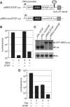
Repression of cap-independent translation of nonSTOP mRNA. (A) Schematic drawing of reporter genes. Open boxes indicate ORFs. Black boxes indicate an internal ribosome entry site (IRES) from encephalomyocarditis virus (ECMV). Shaded boxes indicate a polyadenylation signal. The termination codon of Firefly luciferase gene is indicated by a STOP sign. (B) Luciferase assays were performed following transfection of HeLa cells and Firefly luciferase activities (Luc) were normalized to cotransfected Renilla luciferase activities. Lane 1: pGL STOP; lane 2: pGL nonSTOP; lane 3: pIRES-STOP Luc; lane 4: pIRES-nonSTOP Luc. Error bars represent standard deviations of four independent experiments. (C) Northern blot analysis of 10 μg total RNA prepared from HeLa cells transfected with pGL STOP (lane 1), pGL nonSTOP (lane 2), pIRES-STOP Luc (lane 3), or pIRES-nonSTOP Luc (lane 4) and probed with 32P-Firefly luciferase probe (Luc, upper panel) or 32P-Renilla luciferase probe (RL, lower panel). (D) Transfection of in vitro synthesized RNAs into HeLa cells. Measured Firefly luciferase activities of HeLa cells transfected with Firefly luciferase mRNAs were normalized to the activity of cotransfected Renilla luciferase mRNA. Lane 1: capped wild-type Firefly luciferase mRNA; lane 2: capped nonSTOP Firefly luciferase mRNA; lane 3: uncapped wild-type Firefly luciferase mRNA; lane 4: uncapped nonSTOP Firefly luciferase mRNA. There was no difference in the expression level of cotransfected Renilla mRNA under the four conditions. Error bars represent standard deviations of four independent experiments.
Figure 5
Figure 5
In vitro run-off translation programmed with mRNA associating with polysomes. Polysomal fraction (#13 from Figure 3) containing either wild-type Firefly luciferase (STOP Luc) or nonSTOP Firefly luciferase (nonSTOP Luc) mRNA was prepared by sucrose gradient centrifugation in the absence of cycloheximide, and used to program rabbit reticulocyte lysate at 30°C in the presence of the 50 μg/ml aurintricarboxylic acid, an initiation inhibitor. Small aliquots were removed and luciferase activities were measured at the indicated time. Luciferase activities obtained are presented as the mean values±experimental errors, which was obtained from two independent experiments. Black and red circles represent luciferase activity produced from reactions containing polysome-associated STOP Luc and nonSTOP Luc mRNA, respectively.
Figure 6
Figure 6
Short truncated polypeptides are produced from nonSTOP mRNA in vitro. In vitro synthesized mRNA coding FLAG-tagged Firefly luciferase (STOP Luc) or FLAG-tagged nonSTOP Firefly luciferase (nonSTOP Luc) were incubated at 30°C with rabbit reticulocyte lysate supplemented with 14C-leucine and all necessary components for translation. (A) Small aliquots were sampled and Firefly luciferase activities were measured at the indicated times. Luciferase activities are shown as the mean±experimental error, which was obtained from two independent experiments. Black and red circles represent the result of STOP Luc mRNA and nonSTOP Luc mRNA, respectively. White circles are the results from control translations lacking input mRNA. (B) 14C-label incorporation into acid-insoluble material. Sampled small aliquots were spotted on 3MM paper, and then incubated with 5% trichloroacetic acid solution at 80°C for 30 min, washed, and then quantitated by liquid scintillation counting. Radioactivity counts are shown as the mean values±experimental errors, which were obtained from two independent experiments. Black and red circles represent acid-insoluble 14C-label incorporation produced from STOP Luc mRNA and nonSTOP Luc mRNAs, respectively. White circles represent the result of control reactions lacking mRNA. (C) In vitro translation reactions performed with no mRNA (lane 1), FLAG-Firefly luciferase mRNA (lane 2), or FLAG-nonSTOP Firefly luciferase mRNA (lane 3) were processed for immunoprecipitation using an anti-FLAG antibody (M2 mAb, SIGMA). Immunoprecipitates were analyzed by SDS–PAGE and the signals visualized using a BAS-2500 imaging analyzer.
Figure 7
Figure 7
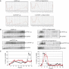
Inhibition of translation in vivo indicates that translating nonSTOP mRNA is distinct from STOP mRNA. (A) HeLa cells transfected with either pGL STOP (top panels) or pGL nonSTOP (bottom panels) were treated with 5 μM hippuristanol for 0 (left panels) or 10 min (right panels) before cell lysis and separation of polyribosomes. Absorbance at 254 nm profiling during fractionation is shown. (B) Collected fractions were subjected to Northern blot analysis with 32P-Luc or 32P-GAPDH probe. STOP Luc indicates signals of wild-type luciferase mRNA expressed from pGL STOP plasmid. NonSTOP Luc indicates signals of nonSTOP luciferase mRNA expressed from pGL nonSTOP plasmid. (C) Luciferase mRNA signals normalized to the total signal across all fractions in Figure 7B (left panel: no hippuristanol; right panel: 5 μM hippuristanol for 10 min). Each value is shown as the mean value±experimental errors, which were obtained from two independent experiments. Black and red circles represent STOP Luc and nonSTOP Luc mRNA, respectively.
Figure 8
Figure 8
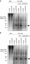
32P-labeled poly(A) fragments protected by ribosomes on nonSTOP mRNA upon micrococcal nuclease (MN) treatment. (A) In vitro synthesized Firefly luciferase mRNA having a 32P-labeled poly(A) tail (STOP Luc+32P-poly(A); lanes 1, 3, 5, and 7) or nonSTOP Firefly luciferase mRNA having a 32P-labeled poly(A) tail (nonSTOP Luc+32P-poly(A); lanes 2, 4, 6, and 8) was incubated with 20 μl rabbit reticulocyte lysate for 60 min at 30°C. Following in vitro translations, either aurintricarboxylic acid (ATA) (50 μM; lanes 5 and 6) or puromycin (200 μM; lanes 7 and 8) were added and then incubated for 60 min. Upon addition of 1 mg/ml cycloheximide and 40 mM CaCl2, the mixture was digested with 30 units micrococcal nuclease for 15 min at 30°C (lanes 3–8). After termination of the nuclease digestion by the addition of 160 mM EGTA, 0.5% SDS and 1 mg/ml proteinase K were added and the mixture was incubated for 30 min at 37°C. RNA was extracted by water-saturated phenol/chloroform and precipitated with ethanol in the presence of 0.2 mg/ml glycogen. The resulting pellet was resuspended in RNA running solution (10 M urea, 1 × TBE and 0.1% BPB) and analyzed by electrophoresis in a 8 M urea, 10% polyacrylamide gel and the signal visualized using a BAS-2500 imaging analyzer. The undigested mRNAs (lanes 1 and 2) represent 1/5th the amount of samples loaded in the other lanes. (B) Examination of the effect of two different initiation inhibitors for the appearance of ribosome-protected poly(A) fragments. Indicated luciferase mRNAs harboring a 32P-labeled poly(A) tail were used to program an in vitro translation reaction, then incubated additionally in the presence of either aurintricarboxylic acid (50 μM; lanes 5 and 6) or hippuristanol (10 μM; lanes 7 and 8). After terminating the reaction by addition of cycloheximide, RNAs were digested and analyzed as mentioned above. The arrows at the right position of panels indicate the position of ∼40 nt poly(A) fragments.

References

    1. Abo T, Inada T, Ogawa K, Aiba H (2000) SsrA-mediated tagging and proteolysis of LacI and its role in the regulation of lac operon. EMBO J 19: 3762–3769 - PMC - PubMed
    1. Amrani N, Sachs MS, Jacobson A (2006) Early nonsense: mRNA decay solves a translational problem. Nat Rev Mol Cell Biol 7: 415–425 - PubMed
    1. Baker KE, Parker R (2004) Nonsense-mediated mRNA decay: terminating erroneous gene expression. Curr Opin Cell Biol 16: 293–299 - PubMed
    1. Behm-Ansmant I, Izaurralde E (2006) Quality control of gene expression: a stepwise assembly pathway for the surveillance complex that triggers nonsense-mediated mRNA decay. Genes Dev 20: 391–398 - PubMed
    1. Bordeleau ME, Mori A, Oberer M, Lindqvist L, Chard LS, Higa T, Belsham GJ, Wagner G, Tanaka J, Pelletier J (2006) Functional characterization of IRESes by an inhibitor of the RNA helicase eIF4A. Nat Chem Biol 2: 213–220 - PubMed

Publication types