. 2007 May 2;26(9):2421-31.
doi: 10.1038/sj.emboj.7601677. Epub 2007 Apr 19.

Structures of modified eEF2 80S ribosome complexes reveal the role of GTP hydrolysis in translocation

Affiliations

Structures of modified eEF2 80S ribosome complexes reveal the role of GTP hydrolysis in translocation

Derek J Taylor et al. EMBO J. .

Abstract

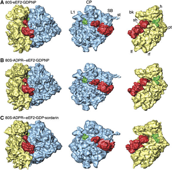
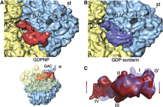
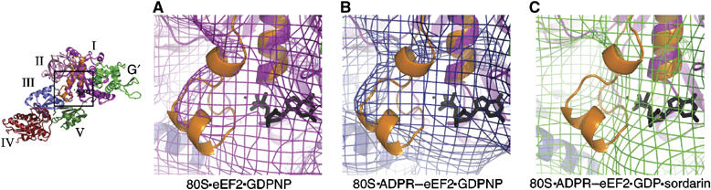
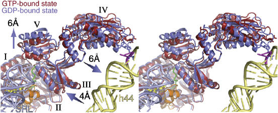
On the basis of kinetic data on ribosome protein synthesis, the mechanical energy for translocation of the mRNA-tRNA complex is thought to be provided by GTP hydrolysis of an elongation factor (eEF2 in eukaryotes, EF-G in bacteria). We have obtained cryo-EM reconstructions of eukaryotic ribosomes complexed with ADP-ribosylated eEF2 (ADPR-eEF2), before and after GTP hydrolysis, providing a structural basis for analyzing the GTPase-coupled mechanism of translocation. Using the ADP-ribosyl group as a distinct marker, we observe conformational changes of ADPR-eEF2 that are due strictly to GTP hydrolysis. These movements are likely representative of native eEF2 motions in a physiological context and are sufficient to uncouple the mRNA-tRNA complex from two universally conserved bases in the ribosomal decoding center (A1492 and A1493 in Escherichia coli) during translocation. Interpretation of these data provides a detailed two-step model of translocation that begins with the eEF2/EF-G binding-induced ratcheting motion of the small ribosomal subunit. GTP hydrolysis then uncouples the mRNA-tRNA complex from the decoding center so translocation of the mRNA-tRNA moiety may be completed by a head rotation of the small subunit.


Figures

Figure 1
Figure 1
Cryo-EM reconstructions of 80S·eEF2/ADPR-eEF2 complexes. The computationally separated densities from the 80S·eEF2·GDPNP (A), the 80S·ADPR-eEF2·GDPNP (B), and the 80S·ADPR-eEF2·GDP·sordarin (C) complexes. The 60S subunit (LSU) is colored blue, the 40S subunit (SSU) is yellow, the P/E-site tRNA is green, and eEF2 and ADPR-eEF2 are red. Density attributed to the ADPR moiety is circled in (B). Landmarks in (A) are: CP, central protuberance; SB, stalk base; st, stalk; L1, L1 protuberance; h, head; bk, beak; pt, point; sh, shoulder; lf, left foot; rf, right foot.
Figure 2
Figure 2
GTP hydrolysis causes a shift of domains I, II, and G′ of ADPR-eEF2 toward the GAC. Both ADPR-eEF2 and the GAC in the LSU undergo significant rearrangements due to GTP hydrolysis, as seen by comparing densities in the GTP-bound (GDPNP, A) and GDP-bound (GDP:sordarin, B) complexes. These conformational changes induce a reorganization of the contacts between the GAC and ADPR-eEF2, which are highlighted in black and white asterisks in the two panels. (C) Overlaying the densities attributed to ADPR-eEF2 from before (red) and after (blue) GTP hydrolysis exposes the conformational changes induced in ADPR-eEF2. This overlay reveals a shift in domains I, II, and G′ of ADPR-eEF2 toward the GAC upon GTP hydrolysis, while domains III, IV, and V are seen to shift in the opposite direction, away from the LSU. DC, decoding center; GAC, GTPase associated center; st, stalk.
Figure 3
Figure 3
Superimposition of the Switch 1 loop from the EF–Tu ternary complex. The Switch 1 loop from the crystal structure of the EF–Tu ternary complex was superimposed onto our quasi-atomic models of eEF2 bound to the ribosome by least-squares alignment of the conserved helix A in domain I of the two structures. The Switch 1 loop from EF–Tu is accommodated by density in the cryo-EM structures of eEF2 in the GTP state (A) and ADPR-eEF2 in the GTP state (B), but the density of ADPR-eEF2 in the GDP state (C) shows that the Switch 1 loop has become disordered after GTP hydrolysis has occurred. Domains of eEF2 and ADPR-eEF2 are color-coded, the Switch 1 loop is colored orange, and GDP is colored black in all three panels. This figure was generated using PyMOL (DeLano, 2002).
Figure 4
Figure 4
Stereo-view of domain movements in ADPR-eEF2 caused by GTP hydrolysis. The real-space refined, quasi-atomic models of ADPR-eEF2 before (red) and after (blue) GTP hydrolysis are shown together. GTP hydrolysis causes a shift in domains III and V of eEF2 away from domain I. Domain IV and V rotate slightly, as a single rigid body, resulting in a 6 Å shift of the tip of domain IV toward helix 44 in the rRNA of the SSU. The proximity of the SRL (light blue) to the Switch 1 loop (orange) in the GTP-bound state of the quasi-atomic model supports the role of the SRL in catalyzing GTP hydrolysis. GDP is shown in yellow stick representation, the Switch 2 loop is colored green, and the Switch 1 loop in the GTP-bound state is orange. Helix 44 from the 18S rRNA of the SSU is colored light yellow and the SRL from the 25S rRNA of the LSU is colored light blue. The location of two universally conserved adenine bases, A1492 and A1493 (E. coli numbering) in helix 44, that compose part of the ribosomal decoding center, is represented as magenta sticks. This figure was generated with PyMOL (DeLano, 2002).
Figure 5
Figure 5
Docking of the X-ray structure of the ADPR moiety into the cryo-EM density map. (A) Comparison of the density obtained from the eEF2 (magenta mesh) and ADPR-eEF2 (blue mesh) maps, both with GDPNP, shows that density attributed to the ADPR occupies the ribosomal A site before GTP hydrolysis. (B) Fitting of the ADPR moiety after GTP hydrolysis in the 80S·ADPR-eEF2·GDP·sordarin cryo-EM density was facilitated by superimposition of domain IV of the ADPR-eEF2·exotoxin A crystal structure (Jorgensen et al, 2005). The docked ADPR moiety is well accounted for by extra density in this map. (C) A comparison of density obtained from the 80S·ADPR-eEF2·GDPNP (blue mesh) and 80S·ADPR-eEF2·GDP·sordarin (green mesh) complexes indicates that the ADPR moiety moves out of the A site, and into helix 44, after GTP hydrolysis. In all panels, ADPR-eEF2 is red, the ADPR moiety is colored according to atom composition, SSU rRNA is yellow, helix 44 of the SSU is orange, with the phosphate backbone of the conserved adenines in helix 44 (A1492 and A1493 in E. coli) colored cyan.
Figure 6
Figure 6
Comparison of ADPR-eEF2 conformational changes due to sordarin binding versus GTP hydrolysis. (A) The computationally separated densities from the 80S·ADPR-eEF2·GDPNP·sordarin complex reveals that the ADPR moiety occupies the intersubunit space (circled), as it does in the 80S·ADPR-eEF2·GDPNP complex (Figure 1B). This observation indicates that the binding of sordarin does not induce conformational changes in eEF2, when bound to the ribosome and GDPNP. (B) The primary difference in density between the 80S·ADPR-eEF2·GDPNP and 80S·ADPR-eEF2·GDPNP·sordarin complexes is density that can be attributed to the presence of sordarin, which is represented as yellow sticks in both panels. (C) Density attributed to ADPR-eEF2 from the 80S·ADPR-eEF2·GDPNP map (red) is virtually superimposable with that from the 80S·ADPR-eEF2·GDPNP·sordarin map (gray). Conversely, superimposition of density attributed to ADPR-eEF2 from the 80S·ADPR-eEF2·GDP·sordarin map (blue) with that from the 80S·ADPR-eEF2·GDPNP·sordarin map (gray) demonstrates significant conformational changes in the factor that are due exclusively to GTP hydrolysis. The direction and magnitude of these conformational changes are in agreement with those in the quasi-atomic models obtained after real-space refinement (D). The quasi-atomic models from the 80S·ADPR-eEF2·GDPNP (red) and 80S·ADPR-eEF2·GDPNP·sordarin (gray) complexes are very similar, whereas a comparison of the 80S·ADPR-eEF2·GDP·sordarin (blue) and 80S·ADPR-eEF2·GDPNP·sordarin (gray) quasi-atomic models shows conformational changes that are a result of GTP hydrolysis.
Figure 7
Figure 7
Head rotation of the 40S subunit of the yeast 80S ribosome. Binding of eEF2 induces a subunit rearrangement that involves ratcheting of the SSU with respect to the LSU, as well as head rotations in the SSU. To discriminate the difference induced exclusively by the head rotation, we superimposed the bodies of the SSU from the quasi-atomic models of the pre-translocational (Spahn et al, 2001) and the ratcheted, eEF2-bound (Spahn et al, 2004) 80S ribosomes, by explicit least-squares fitting using the program O (Jones et al, 1991). The SSU of the pre-translocational ribosome with P-site tRNA (PDB ID: 1K5X) is shown in tan, and that of the eEF2-bound ribosome (PDB ID: 1S1H) is shown in blue. Highlighted residues U955 and A1339 are in regions known to interact with ribosome-bound tRNA (Yusupov et al, 2001). This alignment demonstrates that head rotation alone can account for 12–13 Å movements of tRNA during translocation. Similar ratcheting and head rotations have been noted for the SSU of the bacterial ribosome (Schuwirth et al, 2005), suggesting that the mechanism of translocation is conserved.

References

    1. Aevarsson A, Brazhnikov E, Garber M, Zheltonosova J, Chirgadze Y, al-Karadaghi S, Svensson LA, Liljas A (1994) Three-dimensional structure of the ribosomal translocase: elongation factor G from Thermus thermophilus. EMBO J 13: 3669–3677 - PMC - PubMed
    1. Agrawal RK, Heagle AB, Penczek P, Grassucci RA, Frank J (1999) EF-G-dependent GTP hydrolysis induces translocation accompanied by large conformational changes in the 70S ribosome. Nat Struct Biol 6: 643–647 - PubMed
    1. Agrawal RK, Penczek P, Grassucci RA, Frank J (1998) Visualization of elongation factor G on the Escherichia coli 70S ribosome: the mechanism of translocation. Proc Natl Acad Sci USA 95: 6134–6138 - PMC - PubMed
    1. Allen GS, Zavialov A, Gursky R, Ehrenberg M, Frank J (2005) The cryo-EM structure of a translation initiation complex from Escherichia coli. Cell 121: 703–712 - PubMed
    1. Armstrong S, Yates SP, Merrill AR (2002) Insight into the catalytic mechanism of Pseudomonas aeruginosa exotoxin A. Studies of toxin interaction with eukaryotic elongation factor-2. J Biol Chem 277: 46669–46675 - PubMed

Publication types

MeSH terms