. 2007 Apr;3(4):e57.
doi: 10.1371/journal.ppat.0030057.

Staphylococcal biofilm exopolysaccharide protects against Caenorhabditis elegans immune defenses

Affiliations

Staphylococcal biofilm exopolysaccharide protects against Caenorhabditis elegans immune defenses

Jakob Begun et al. PLoS Pathog. 2007 Apr.

Abstract

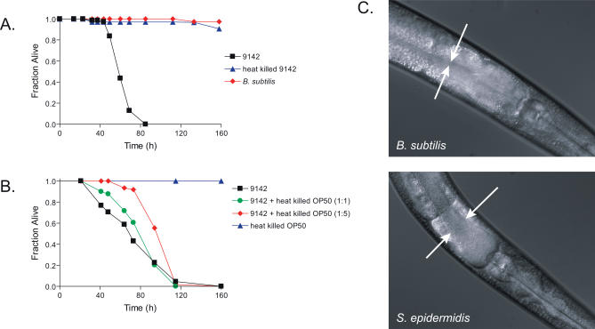
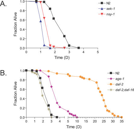
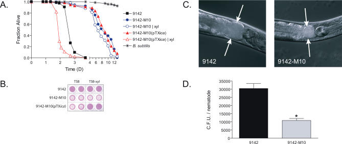
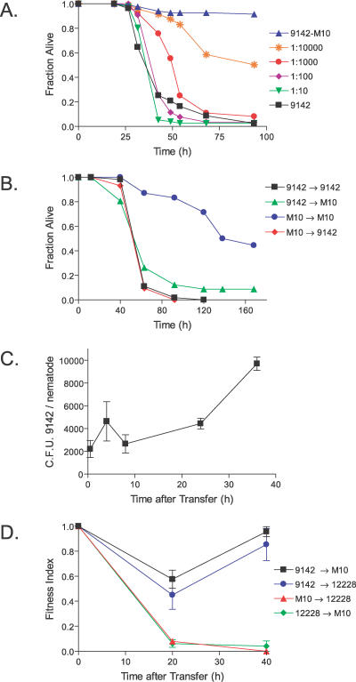
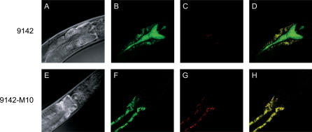
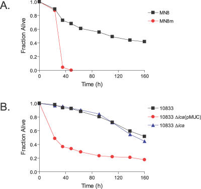
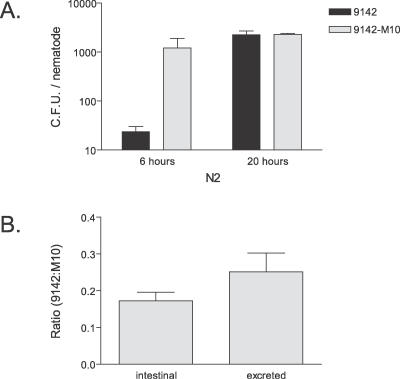
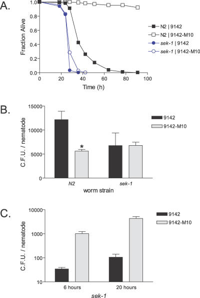
Staphylococcus epidermidis and Staphylococcus aureus are leading causes of hospital-acquired infections that have become increasingly difficult to treat due to the prevalence of antibiotic resistance in these organisms. The ability of staphylococci to produce biofilm is an important virulence mechanism that allows bacteria both to adhere to living and artificial surfaces and to resist host immune factors and antibiotics. Here, we show that the icaADBC locus, which synthesizes the biofilm-associated polysaccharide intercellular adhesin (PIA) in staphylococci, is required for the formation of a lethal S. epidermidis infection in the intestine of the model nematode Caenorhabditis elegans. Susceptibility to S. epidermidis infection is influenced by mutation of the C. elegans PMK-1 p38 mitogen-activated protein (MAP) kinase or DAF-2 insulin-signaling pathways. Loss of PIA production abrogates nematocidal activity and leads to reduced bacterial accumulation in the C. elegans intestine, while overexpression of the icaADBC locus in S. aureus augments virulence towards nematodes. PIA-producing S. epidermidis has a significant survival advantage over ica-deficient S. epidermidis within the intestinal tract of wild-type C. elegans, but not in immunocompromised nematodes harboring a loss-of-function mutation in the p38 MAP kinase pathway gene sek-1. Moreover, sek-1 and pmk-1 mutants are equally sensitive to wild-type and icaADBC-deficient S. epidermidis. These results suggest that biofilm exopolysaccharide enhances virulence by playing an immunoprotective role during colonization of the C. elegans intestine. These studies demonstrate that C. elegans can serve as a simple animal model for studying host-pathogen interactions involving staphylococcal biofilm exopolysaccharide and suggest that the protective activity of biofilm matrix represents an ancient conserved function for resisting predation.


Conflict of interest statement

Competing interests. The authors have declared that no competing interests exist.

Figures

Figure 1
Figure 1. S. epidermidis Kills C. elegans and Causes Intestinal Distension
(A) C. elegans killing assays on lawns of live S. epidermidis 9142 (squares), heat-killed S. epidermidis 9142 (triangles), or B. subtilis strain RL2244 (diamonds). (B) Survival of C. elegans exposed to live S. epidermidis 9142 (squares), admixtures of live S. epidermidis 9142 and heat-killed E. coli OP50 at a ratio of 1:1 (circles) or 1:5 (diamonds), or heat-killed E. coli OP50 alone (triangles). (C) N2 C. elegans were exposed to B. subtilis RL2244 or S. epidermidis 9142 for 24 h and then visualized by Nomarski differential contrast microscopy. Arrows demarcate the intestinal tract lumen. Magnification, ×40.
Figure 2
Figure 2. C. elegans Mutants with Altered Immune Function Show Differential Sensitivity to S. epidermidis
(A) Survival of wild-type N2 C. elegans (squares) compared to survival of the immunocompromised, p38 MAP kinase pathway mutants sek-1(ag1) (triangles, p < 0.0001) or nsy-1(ag3) (inverted triangles, p < 0.0001) when exposed to S. epidermidis 9142. (B) Survival of N2 animals (squares) compared to survival of the pathogen-resistant, insulin signaling pathway mutants age-1(hx546) (diamonds, p < 0.0001) or daf-2(e1370) (circles, p < 0.0001) when exposed to S. epidermidis 9142. Survival of daf-2(e1370);daf-16(mgDf47) mutants (asterisks) demonstrates that daf-16(mgDf47) suppresses daf-2(e1370) enhanced pathogen resistance.
Figure 3
Figure 3. S. epidermidis Virulence in the C. elegans Infection Model Depends on icaADBC
(A) Survival of C. elegans infected with S. epidermidis strain 9142-M10 (circles), which contains a transposon insertion in the icaA gene, and the complemented strain 9142-M10(pTXica) (triangles), which carries the icaADBC operon driven by the xylose-inducible PxylA promoter, compared to wild-type S. epidermidis 9142 (squares) and B. subtilis (asterisks). Survival assays were performed under standard conditions (closed symbols) or using plates supplemented with 2% xylose (xyl, open symbols). (B) Biofilm formation of S. epidermidis 9142, 9142-M10, and 9142-M10(pTXica) on polystyrene. Attachment to polystyrene 96-well flat bottom microtiter plate was performed as described in the Materials and Methods. Strains were grown in TS broth without supplementation (TSB) or supplemented with 2% xylose (TSB-xyl). (C) Nomarski micrograph of C. elegans after feeding for 24 h on S. epidermidis 9142 or 9142-M10. Arrows demarcate the intestinal tract lumen. Magnification, ×40. (D) Quantification of intestinal bacteria obtained by disruption of worms after 24 h of feeding on either S. epidermidis 9142 or 9142-M10. Values represent the mean of five samples with approximately eight worms per sample ± standard error of the mean (SEM). The asterisk indicates a significant difference (p < 0.001).
Figure 4
Figure 4. The Fitness of Intraluminal S. epidermidis during Colonization of C. elegans Is icaADBC-Dependent
(A) Survival of nematodes feeding on mixed lawns of S. epidermidis 9142 and 9142-M10 in the ratios of 9142:9142-M10 indicated. (B) C. elegans exposed to S. epidermidis 9142 for 12 h and then transferred to S. epidermidis 9142-M10 (triangles) die with similar kinetics to worms transferred from 9142 to 9142 (squares) or 9142-M10 to 9142 (diamonds) and much more rapidly than control worms transferred from 9142-M10 to 9142-M10 (circles). (C) Intestinal proliferation of S. epidermidis 9142 over time in C. elegans feeding on 9142 for 12 h and transferred to 9142-M10. Values represent the mean of three samples with approximately ten worms per sample ± SEM. (D) Nematodes exposed to S. epidermidis 9142 for 12 h and transferred either to 9142-M10 (squares) or a second ica-deficient S. epidermidis strain ATCC 12228 (circles) retain a high proportion of 9142 in their intestinal tracts, whereas those exposed first to the ica-deficient 9142-M10 and transferred to ica-deficient ATCC 12228 (triangles), or vice versa (diamonds), do not retain the initial bacteria in their digestive tracts. Fitness Index is defined as: (pulse S. epidermidis strain C.F.U.) / (pulse S. epidermidis strain C.F.U. + chase S. epidermidis strain C.F.U.). Values represent the mean of three samples with approximately ten worms per sample ± SEM.
Figure 5
Figure 5. Production of PIA by S. epidermidis within the C. elegans Intestinal Tract
Confocal microscopy images of C. elegans feeding on S. epidermidis labeled with FITC-conjugated WGA lectin, which selectively labels the exopolysaccharide of the biofilm matrix. Nematodes feeding on labeled wild-type S. epidermidis 9142 (A–D) or 9142-M10 (E–H). (A and E) Nomarski differential interference contrast image of anterior portion of nematodes. (B and F) Green fluorescence due to FITC-labeled lectin adhering to S. epidermidis exopolysaccharide and C. elegans intestinal autofluorescence. (C and G) Red fluorescence resulting from intestinal autofluorescence. (D and H) Merged fluorescent images in which green demonstrates bound lectin and yellow demonstrates intestinal autofluorescence.
Figure 6
Figure 6. Overexpression of icaADBC in S. aureus Enhances Virulence
(A) Survival of C. elegans N2 infected by the icaADBC-overexpressing S. aureus strain MN8m (circles) compared to wild-type MN8 (squares) (p < 0.0001). (B) Survival of C. elegans N2 infected by S. aureus strain 10833 (squares), 10833 Δica (triangles, p > 0.05), and by strain 10833 Δica(pMUC) (circles, p < 0.0001).
Figure 7
Figure 7. Intestinal and Fecal Ratios of Wild-Type and Biofilm-Deficient S. epidermidis in C. elegans when Fed Mixed Lawns
(A) Intestinal load of S. epidermidis 9142 (dark bars) and 9142-M10 (light bars) within wild-type N2 C. elegans feeding on mixed lawns in a ratio of 1:100. Bacterial loads were determined in triplicate from approximately ten disrupted worms. Data represent mean ± SEM. (B) Ratio of S. epidermidis 9142 to 9142-M10 of excreted and intestinal bacteria. Colonized N2 C. elegans were allowed to feed on mixed lawns of 9142 and 9142-M10 (ratio 1:100) for 16 h, washed, and then transferred to M9 buffer, in which bacteria were freely excreted. Bacterial loads were determined in triplicate from the homogenates and expelled collections of approximately ten worms. Data represent mean ± SEM.
Figure 8
Figure 8. Immunocompromised C. elegans Are Hypersusceptible to both Wild-Type and Biofilm-Deficient S. epidermidis and Accumulate both Strains Equally
(A) sek-1(km4) mutant worms (circles) are similarly susceptible to both S. epidermidis 9142 (closed symbols) and 9142-M10 (open symbols), whereas wild-type N2 nematodes (squares) are differentially resistant to 9142-M10 (p < 0.0001). (B) S. epidermidis 9142 (solid bars) is better able to colonize N2 worms than 9142-M10 (open bars), but there is no difference between colonization rates in sek-1(km4) mutant nematodes after 16 h of feeding on S. epidermidis. Data represent mean ± SEM. The asterisk indicates a significant difference (p < 0.05). (C) Intestinal load of S. epidermidis 9142 (dark bars) and 9142-M10 (light bars) within sek-1(km4) C. elegans feeding on mixed lawns in a ratio of 1:100. Bacterial loads were determined in triplicate from approximately ten disrupted worms. Data represent mean ± SEM.

References

    1. Costerton JW, Stewart PS, Greenberg EP. Bacterial biofilms: A common cause of persistent infections. Science. 1999;284:1318–1322. - PubMed
    1. Mah TF, O'Toole GA. Mechanisms of biofilm resistance to antimicrobial agents. Trends Microbiol. 2001;9:34–39. - PubMed
    1. Mack D, Fischer W, Krokotsch A, Leopold K, Hartmann R, et al. The intercellular adhesin involved in biofilm accumulation of Staphylococcus epidermidis is a linear β-1,6-linked glucosaminoglycan: Purification and structural analysis. J Bacteriol. 1996;178:175–183. - PMC - PubMed
    1. Mack D, Nedelmann M, Krokotsch A, Schwarzkopf A, Heesemann J, et al. Characterization of transposon mutants of biofilm-producing Staphylococcus epidermidis impaired in the accumulative phase of biofilm production: Genetic identification of a hexosamine-containing polysaccharide intercellular adhesin. Infect Immun. 1994;62:3244–3253. - PMC - PubMed
    1. Götz F. Staphylococcus and biofilms. Mol Microbiol. 2002;43:1367–1378. - PubMed

Publication types

MeSH terms

Substances

LinkOut - more resources