. 2007 Jun 22;1154(1-2):308-18.
doi: 10.1016/j.chroma.2007.03.126. Epub 2007 Apr 6.

Elution of tightly bound solutes from concanavalin A Sepharose. Factors affecting the desorption of cottonmouth venom glycoproteins

Affiliations

Elution of tightly bound solutes from concanavalin A Sepharose. Factors affecting the desorption of cottonmouth venom glycoproteins

Anastasiya S Soper et al. J Chromatogr A. .

Abstract

Some glycoproteins bind so tightly to concanavalin A Sepharose that common desorption techniques are ineffective, so a systematic exploration of factors affecting desorption of cottonmouth venom glycoproteins was undertaken. Glycoprotein desorption is greatly improved by introducing up to four pauses of 5-10 min duration into the elution step. Eluent concentrations above 250 mM methylglucoside or 500 mM methyl-mannoside reduced glycoprotein desorption. Eluent NaCl diminished glycoprotein desorption. Most venom glycoproteins desorb more readily as pH diminishes from 6.0 to 4.0, but phosphodiesterase shows the opposite pattern. Eluents recommended by the supplier for desorbing solutes or for column cleaning were ineffectual.


Figures

Fig. 1
Fig. 1
Glycoprotein elution profiles with 0-4 pauses introduced during desorption. The 1 mL Con A Sepharose column was equilibrated in 50 mM acetic acid/NaOH/500 mM NaCl/1 mM MnCl2/1 mM CaCl2/1 mM MgCl2 (pH 6.0) and desorbed with 100 mM acetic acid/NaOH/1 M methyl-α-d-glucopyranoside (pH 4.0). A short, narrow depression preceding a peak indicates the point at which a 10-min pause was introduced (4, 8, 12, and 16 mL after desorption onset). In all experiments, 20 column volumes (20 mL) of eluent were used.
Fig. 2
Fig. 2
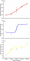
Influence of pause number on glycoprotein and PDE desorption. Glycoprotein desorption increases significantly in sigmoidal fashion with the number of 10-min desorption pauses employed during elution (p < 0.0001, Kruskal-Wallis nonparametric ANOVA). The A280 curve and the results of Bradford and PDE assays suggest that additional pauses (>4) could still desorb additional material, a result also suggested by Fig. 1. In all experiments, 20 column volumes (20 mL) of eluent were used.
Fig. 3
Fig. 3
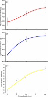
Influence of pause length on glycoprotein and PDE desorption. Glycoprotein desorption is significantly enhanced by increasing pause length in a two-pause model (p = 0.0242, ANOVA). It is unclear whether the curve shape is sigmoidal or hyperbolic; however, pauses longer than 10 min yield diminishing amounts of bound glycoprotein, suggesting that there is little value in using pauses longer than 20 min. It is probably more effective to increase the number rather than the length of pauses. In all experiments, 20 column volumes (20 mL) of eluent were used.
Fig. 4
Fig. 4
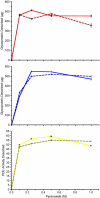
Influence of concentration and type of competitive desorbent. Counterintuitively, desorption of cottonmouth venom glycoproteins with methyl-α-d-glucopyranoside and methyl-α-d-mannopyranoside is generally maximal at 250 mM and 500 mM, respectively. Further increases in pyranoside concentration are counterproductive. PDE desorption is maximal at 500 mM methyl-α-d-glucopyranoside, a little later than for most other venom glycoproteins. Methyl-α-d-glucopyranoside is at least as effective a desorbent for cottonmouth venom glycoproteins as methyl-α-d-mannopyranoside.
Fig. 5
Fig. 5
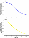
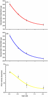
Influence of eluent NaCl concentration on the Bradford and PDE assays. Both assays are negatively impacted by increasing sample NaCl concentrations, as determined from positive controls (p < 0.0001, Kruskal-Wallis nonparametric ANOVA). Data in Fig. 6 were corrected for these effects.
Fig. 6
Fig. 6
Influence of eluent NaCl concentration on glycoprotein and PDE desorption. Increasing eluent NaCl concentrations reduces glycoprotein desorption from Con A Sepharose even after the Bradford and PDE assays are corrected for direct effects on the assay [280 nm absorbance: p < 0.0001, ANOVA; Bradford, p < 0.0001, (Kruskal-Wallis nonparametric ANOVA); PDE: p < 0.0001, (Kruskal-Wallis nonparametric ANOVA)].
Fig. 7
Fig. 7
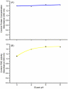
Influence of eluent pH on the Bradford and PDE assays. Across the range of mildly to strongly acidic pH employed in this study, there was no discernible effect of sample pH on the Bradford assay, which employs acidic conditions to facilitate Coomassie dye binding. The slope of the curve deviates significantly from zero (p < 0.0001); however, the goodness of fit is low (r2 = 0.240). PDE, which has a pH optimum close to 9.0, was inhibited by low sample buffer pH. The curve appears hyperbolic here because the pKa of acetic acid in the sample buffer is 4.75. At pH 6.0 acetic acid has little buffering capacity, so above pH 5.0 the sample buffer has a negligible effect on the PDE assay.
Fig. 8
Fig. 8
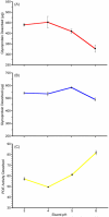
Influence of pH on venom glycoprotein and PDE desorbed from Con A Sepharose. Eluents consisted of 100 mM acetic acid/NaOH/1 M methyl-α-d-glucopyranoside titrated with NaOH to pH 3.0, 4.0, 5.0, or 6.0. Bradford data and PDE activities have been corrected for the effects of pH and ionic strength so that assay data shown here represent only the quantities of glycoprotein and PDE, respectively, eluted from the column (see Fig. 7). The 280 nm absorbance suggests that in general, more glycoprotein is desorbed at lower pH, with no improvement in yield below pH 4.0 (p = 0.0011, Kruskal-Wallis nonparametric ANOVA). This pattern is consistent with supplier literature. The Bradford assay provides a slightly different picture at lower pH, but also suggests that significantly less glycoprotein is desorbed at more neutral pH (p = 0.0001, Kruskal-Wallis nonparametric ANOVA). In contrast, venom PDE desorbs more completely at higher pH, its elution curve being almost a mirror image of the 280 nm curve for all venom glycoproteins (p = 0.0001, Kruskal-Wallis nonparametric ANOVA).
Fig. 9
Fig. 9
Influence of auxiliary and alternative eluents on glycoprotein and PDE desorption from Con A Sepharose. Auxiliary eluents were added to the standard 100 mM acetic acid/NaOH/1 M methyl-α-d-glucopyranoside (pH 4.0) buffer. These included 20% ethylene glycol, 4 M urea, and 40% 2-propanol. Alternative eluents included 100 mM acetic acid/NaOH/50% ethylene glycol (pH 4.0), 100 mM boric acid/NaOH (pH 6.50), 20% ethanol in water and 0.1% Triton X-100. Absorbance (280 nm) and the Bradford assay provide generally similar pictures of glycoprotein desorption, so only the Bradford data are shown here. With the exception of 4 M urea, none of the auxiliary or alternative eluents appeared to be as effective as the standard conditions; however only 40% Propanol and borate were significantly less effective (Bradford assay, p < 0.01, p < 0.001, Kruskal-Wallis nonparametric ANOVA, respectively). The amount of glycoprotein desorbed with 4 M urea was significantly greater than that desorbed with all other eluents except for the standard buffer (Bradford assay, p < 0.001, Kruskal-Wallis nonparametric ANOVA); however, 4 M urea also desorbed Con A itself (Fig. 10).
Fig. 10
Fig. 10
Effects of 4 M urea on Con A Sepharose. A virgin Con A Sepharose column (column filters replaced) was subjected to a standard chromatographic experiment with 100 mM acetic acid/NaOH/1 M methyl-α-d-glucopyranoside/4 M urea (pH 4.0) as the eluent. Sample was never injected onto this column. Con A subunits were released from the resin. Note the reduction in Con A released by a second experiment. SDS PAGE of the released material showed major bands identical to those previously reported for Con A and its naturally occurring fragments [1,18].

Similar articles

Cited by

References

    1. Fountoulakis M, Juranville JF. J. Biochem. Biophys. Methods. 1993;27:127. - PubMed
    1. Hayman MJ, Crumpton MJ. Biochem. Biophys. Res. Commun. 1972;47:923. - PubMed
    1. Bishayee S, Farooqui AA, Bachhawat BK. Indian J. Biochem. Biophys. 1973;10:1. - PubMed
    1. Davey MW, Huang JW, Sulkowski E, Carter WA. J. Biol. Chem. 1974;249:6354. - PubMed
    1. Anderson JC. Biochim. Biophys. Acta. 1975;379:444. - PubMed

Publication types

MeSH terms

LinkOut - more resources