. 2007 Aug 1;364(2):475-85.
doi: 10.1016/j.virol.2007.03.024. Epub 2007 Apr 20.

Characterization of a baculovirus lacking the DBP (DNA-binding protein) gene

Affiliations

Characterization of a baculovirus lacking the DBP (DNA-binding protein) gene

Adam L Vanarsdall et al. Virology. .

Abstract

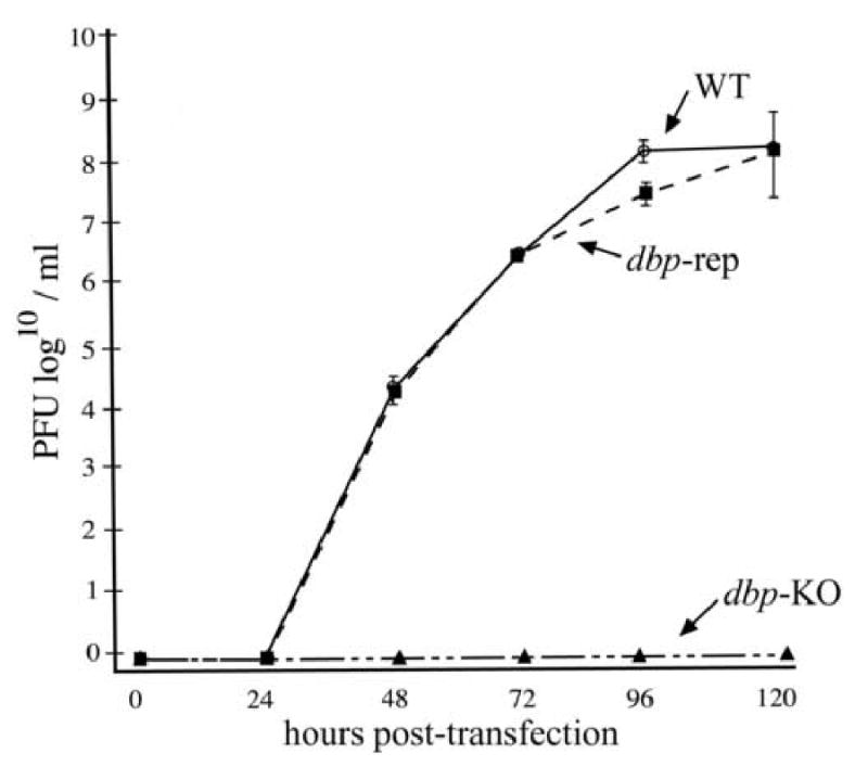
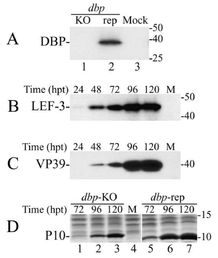
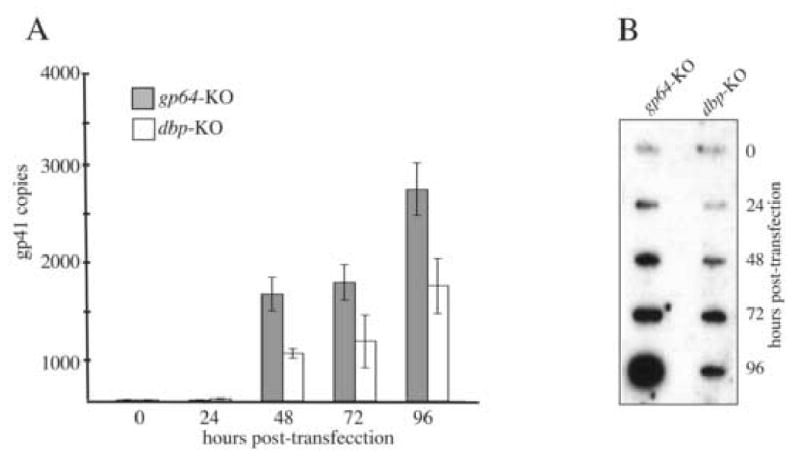
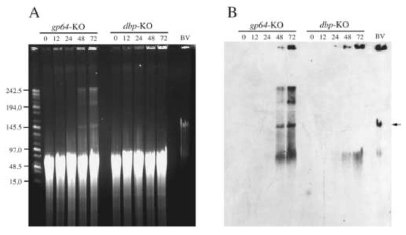
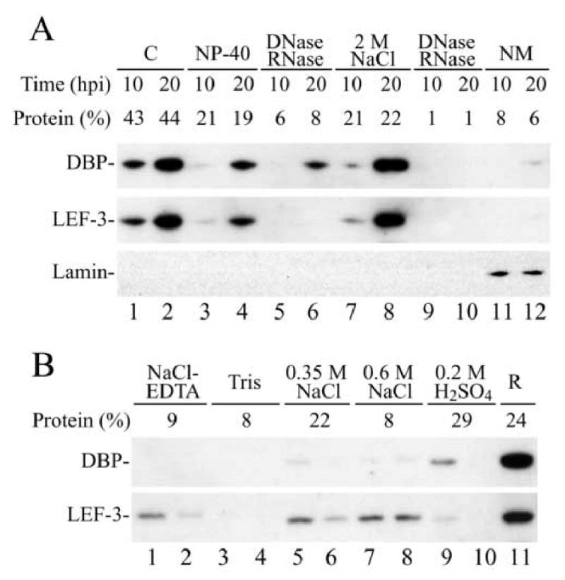
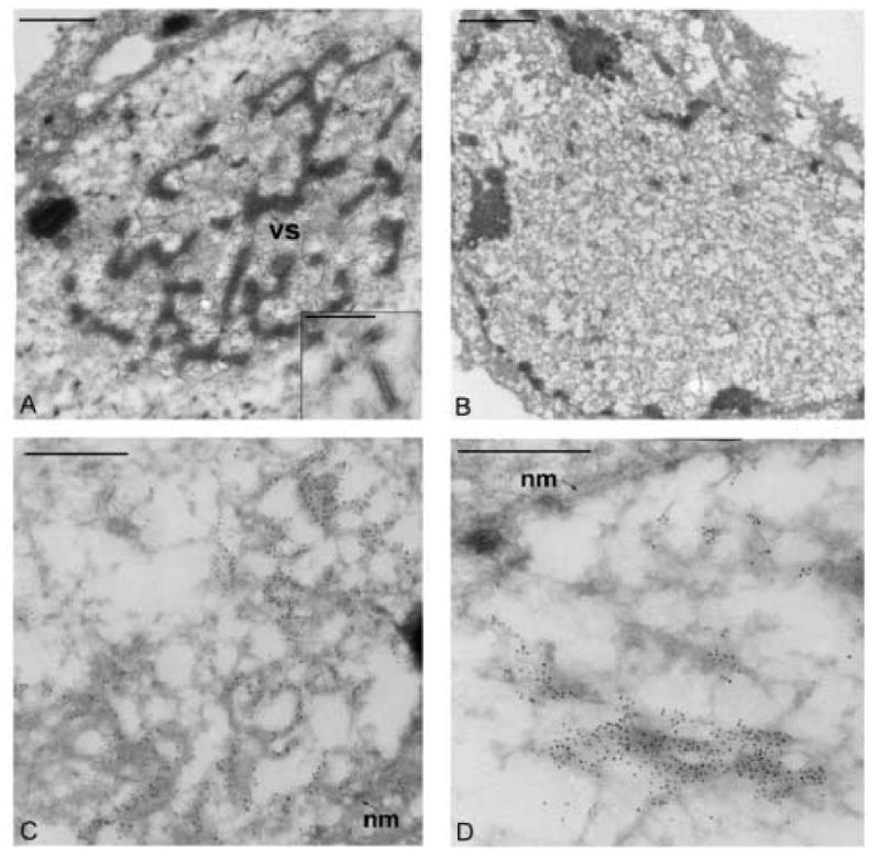
Autographa californica multiple nucleopolyhedrovirus (AcMNPV) encodes two proteins that possess properties typical of single-stranded DNA-binding proteins (SSBs), late expression factor-3 (LEF-3), and a protein referred to as DNA-binding protein (DBP). Whereas LEF-3 is a multi-functional protein essential for viral DNA replication, transporting helicase into the nucleus, and forms a stable complex with the baculovirus alkaline nuclease, the role for DBP in baculovirus replication remains unclear. Therefore, to better understand the functional role of DBP in viral replication, a DBP knockout virus was generated from an AcMNPV bacmid and analyzed. The results of a growth curve analysis indicated that the dbp knockout construct was unable to produce budded virus indicating that dbp is essential. The lack of DBP does not cause a general shutdown of the expression of viral genes, as was revealed by accumulation of early (LEF-3), late (VP39), and very late (P10) proteins in cells transfected with the dbp knockout construct. To investigate the role of DBP in DNA replication, a real-time PCR-based assay was employed and showed that, although viral DNA synthesis occurred in cells transfected with the dbp knockout, the levels were less than that of the control virus suggesting that DBP is required for normal levels of DNA synthesis or for stability of nascent viral DNA. In addition, analysis of the viral DNA replicated by the dbp knockout by using field inversion gel electrophoresis failed to detect the presence of genome-length DNA. Furthermore, analysis of DBP from infected cells indicated that similar to LEF-3, DBP was tightly bound to viral chromatin. Assessment of the cellular localization of DBP relative to replicated viral DNA by immunoelectron microscopy indicated that, at 24 h post-infection, DBP co-localized with nascent DNA at distinct electron-dense regions within the nucleus. Finally, immunoelectron microscopic analysis of cells transfected with the dbp knockout revealed that DBP is required for the production of normal-appearing nucleocapsids and for the generation of the virogenic stroma.


Figures

Fig. 1
Fig. 1
Analysis of budded virus production from bacmid transfected cells. At the indicated time-points, the supernatants from Sf-9 cells transfected with equimolar amounts of the dbp knockout bacmid (dbp-KO), the dbp repair bacmid (dbp-rep), or a control bacmid (AcGUS) were removed and the titers determined by TCID50 end-point dilution assays. The points indicate averages from transfections performed in triplicate and error bars represent standard deviations.
Fig. 2
Fig. 2
Western blot analysis of the synthesis of viral proteins in Sf-9 cells transfected with the dbp knockout (dbp-KO) bacmid. (A) The cells were transfected with the dbp-KO bacmid (lane 1), the repair (rep) bacmid (lane 2), or were mock transfected (lane 3) and harvested at 72 h.p.t. Portions from the cell extracts that corresponded to 0.5 × 10⁴ cells were analyzed by SDS-15% PAGE. The blot was developed with polyclonal antibodies to DBP. (B-C) Time-course of the synthesis of LEF-3 and VP39 in Sf-9 cells transfected with the dbp-KO bacmid. The transfected cells were harvested at the times indicated above the respective lanes. Portions of cell extracts that correspond to 105 cells were subjected to SDS-12% PAGE. The blot was first developed with polyclonal antibodies to LEF-3 (B), and then with monoclonal antibody to VP39 (C). (D) Time-course of the synthesis of P10 in Sf-9 cells transfected with the dbp-KO bacmid (lanes 1-3) or with the repair bacmid (lanes 5-7). The transfected cells were harvested at times indicated above the respective lanes. Portions from the cell extracts that corresponded to 1 ×10⁵ cells were subjected to SDS-15% PAGE. The gel was stained with Coomassie brilliant blue. Lanes “M” show the extract from mock transfected cells. Protein standards (in kDa) are shown on the right.
Fig. 3
Fig. 3
Analysis of viral DNA replication. (A) The bar graph represents the amount of nascent viral DNA synthesized in Sf-9 cells transfected with either the gp64 knockout virus serving as the non-infectious control or the dbp knockout virus. At the designated time-point, total DNA was isolated, treated with DpnI enzyme to digest input bacmid DNA, and analyzed by real-time PCR using a taqman probe and primer set that amplifies a 100 bp region of the AcMNPV gp41 open reading frame. The values displayed are the averages from transfections performed in triplicate with error bars indicating the standard deviations. (B) Slot-blot analysis of replicated viral DNA. At the indicated time-points, one well of a six-well plate of Sf-9 cells transfected with either the gp64 or dbp knockout virus was harvested at the indicated time points in 1 ml PBS and 100 μl of the cell suspension was removed, processed, and applied to nylon membrane using a slot-blot apparatus. The membrane was probed with 32P-labeled AcMNPV genomic DNA.
Fig. 4
Fig. 4
Field-inversion gel electrophoresis of replicated viral DNA. Total DNA was analyzed from Sf-9 cells transfected with either the gp64 knockout serving as the control (gp64-KO) or the dbp knockout (dbp-KO) as described in Material and Methods. Panel A represents the ethidium bromide stained agarose gel and panel B shows the results the Southern blot after hybridization with labeled AcMNPV bacmid DNA. The bacmid contructs that were used for transfection and the time samples were harvested post-transfection are indicated at the top of the panels. The size of the DNA marker in kilo-bases is shown on the left of panel A. “BV” represents viral DNA extracted and processed from extra-cellular budded virus. All samples were digested with Eco81I which cuts the genome at a single site prior to electrophoresis. The arrow indicates the position of full length genomic DNA.
Fig. 5
Fig. 5
Western blot analysis of the association of DBP and LEF-3 with subnuclear structures. (A) AcMNPV-infected Sf-9 cells were harvested at 10 or 20 h.p.i. and homogenized in a hypotonic buffer (see Materials and Methods). Nuclei were separated from the cytoplasmic fraction “C” (lanes 1 and 2) by centrifugation and were sequentially washed with buffer containing 1% Nonidet P-40 (lanes 3 and 4), treated with DNase and RNase (lanes 5 and 6), extracted with buffer containing 2 M NaCl (lanes 7 and 8), treated again with DNase and RNase (lanes 9 and 10), and finally the nuclear matrix “NM” was collected (lanes 11 and 12). A portion from each fraction that corresponded to 0.9 × 10⁵ cells was analyzed by SDS-11% PAGE. The blot was first developed with polyclonal antibodies to DBP, then with polyclonal antibodies to LEF-3, and finally with monoclonal antibody to Drosophila lamin. The amount of protein in each fraction was estimated by optical densitometry of the Coomassie stained gel and is shown as the percentage of total cellular protein above the respective lane. (B) Western blot analysis of the association of DBP and LEF-3 with chromatin structures. Nuclei were isolated from AcMNPV-infected Sf-9 cells harvested at 20 h.p.i. as described in the Materials and Methods and were sequentially extracted twice with buffers containing 75 mM NaCl, 25 mM EDTA (lanes 1 and 2), 10 mM Tris, pH 8.0 (lanes 3 and 4), 0.35 M NaCl (lanes 5 and 6), 0.6 M NaCl (lanes 7 and 8), and with 0.2 M H2SO4 (lanes 9 and 10). Lane R presents the nuclear residue. A portion from each fraction that corresponded to 1.2 × 10⁵ cells was analyzed by SDS-11% PAGE. The blot was first developed with polyclonal antibodies to DBP, and then with polyclonal antibodies to LEF-3. Percentage of nuclear protein extracted by each solution is shown above the lanes.
Fig. 6
Fig. 6
Electron microscopic analysis of thin sections from Sf-9 transfected cells stained with a VP39 antibody. (A) Image of a cell transfected with the gp64-KO control bacmid showing the electron dense virogenic stroma (VS) within the nucleus. The inset panel shows a higher magnification of nucleocapsids immuno-stained with the monoclonal antibody to the major capsid protein VP39. (B) Image of a cell transfected with the dbp-KO bacmid construct showing the lack of an organized virogenic stroma (C and D). Images of a portion of the nucleus from cells transfected with the dbp-KO bacmid construct. The gold label can be seen localized to regions near the nuclear membrane (nm). The bars in panels A and B represent 1 μm and the bars in panels C, D, and the inset represent 0.5 μm.
Fig. 7
Fig. 7
Electron microscopic analysis of Sf-9 cells infected with a DBP repair virus and treated with BrdU. (A-D) Images showing portions of the nucleus from Sf-9 cells at 24 h.p.i. Thin sections were co-stained with a monoclonal antibody to BrdU (20 nm gold) and a polyclonal antibody to DBP (10 nm gold). The bars represent 0.5 μm.

Similar articles

Cited by

References

    1. Braunagel SC, Parr R, Belyavskyi M, Summers MD. Autographa californica nucleopolyhedrovirus infection results in Sf9 cell cycle arrest at G2/M phase. Virology. 1998;244:195–211. - PubMed
    1. Busch H. Ubiquitination of proteins. Methods Enzymol. 1984;106:238–62. - PubMed
    1. Campbell MJ. Lipofection reagents prepared by a simple ethanol injection technique. Biotechniques. 1995;18:1027–32. - PubMed
    1. Cunningham RP, Berger H. Mutations affecting genetic recombination in bacteriophage T4D. I. Pathway analysis. Virology. 1977;80:67–82. - PubMed
    1. Datsenko KA, Wanner BL. One-step inactivation of chromosomal genes in Escherichia coli K-12 using PCR products. Proc Natl Acad Sci U S A. 2000;97:6640–5. - PMC - PubMed

Publication types

MeSH terms