

Cell autonomous requirement for TGF-beta signaling during odontoblast differentiation and dentin matrix formation
- PMID: 17449229
- PMCID: PMC2704601
- DOI: 10.1016/j.mod.2007.02.003
Cell autonomous requirement for TGF-beta signaling during odontoblast differentiation and dentin matrix formation
Abstract
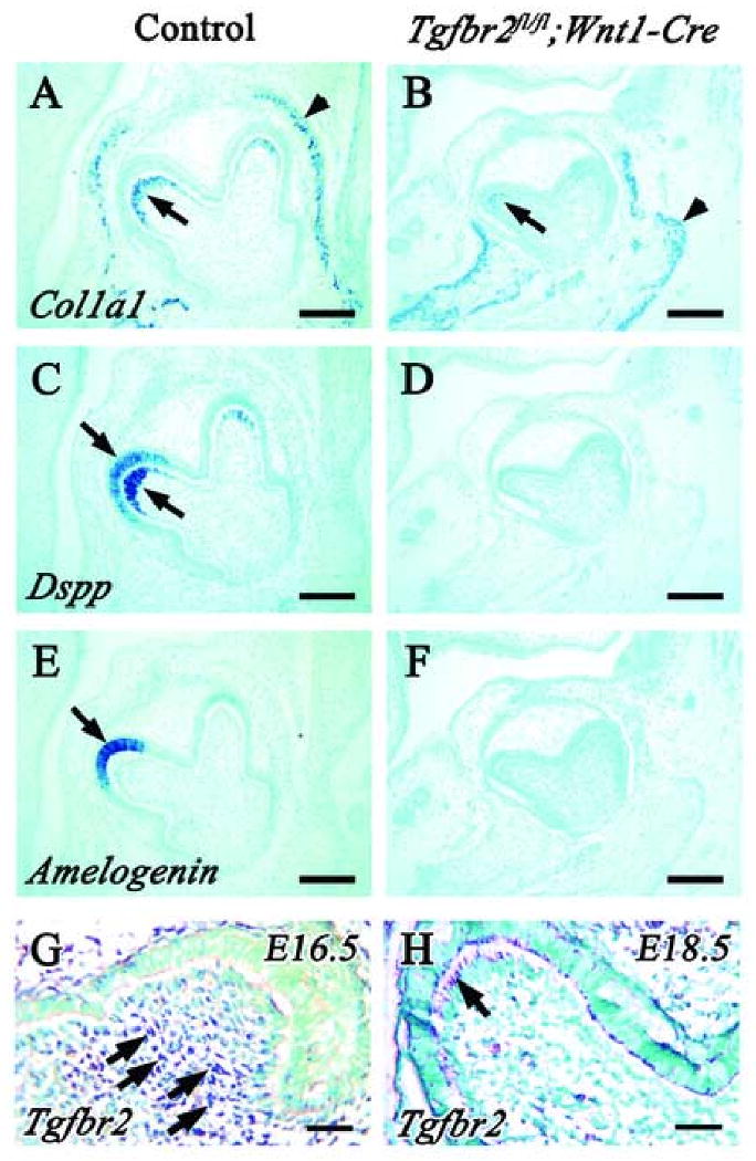
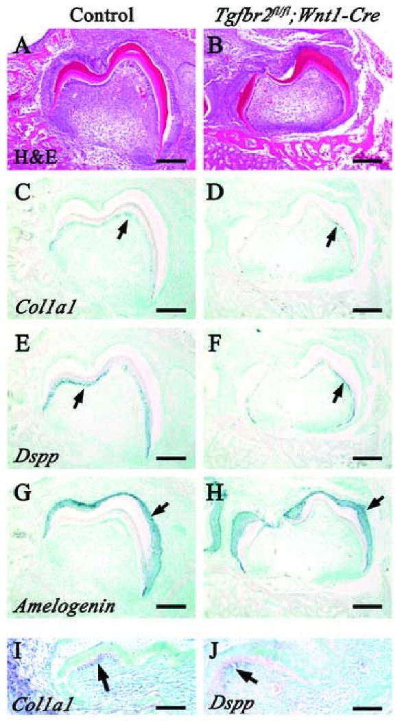
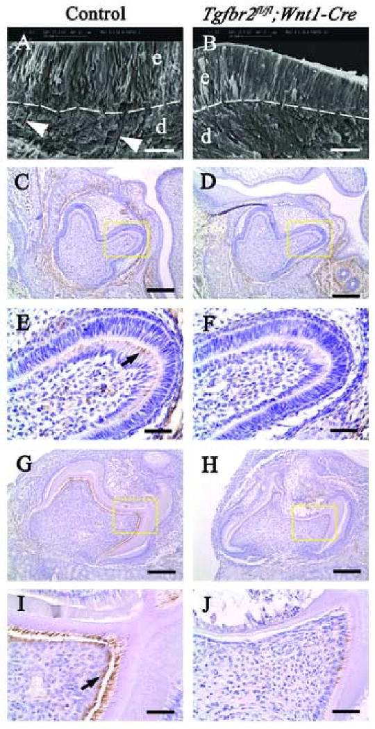
TGF-beta subtypes are expressed in tissues derived from cranial neural crest cells during early mouse craniofacial development. TGF-beta signaling is critical for mediating epithelial-mesenchymal interactions, including those vital for tooth morphogenesis. However, it remains unclear how TGF-beta signaling contributes to the terminal differentiation of odontoblast and dentin formation during tooth morphogenesis. Towards this end, we generated mice with conditional inactivation of the Tgfbr2 gene in cranial neural crest derived cells. Odontoblast differentiation was substantially delayed in the Tgfbr2(fl/fl);Wnt1-Cre mutant mice at E18.5. Following kidney capsule transplantation, Tgfbr2 mutant tooth germs expressed a reduced level of Col1a1 and Dspp and exhibited defects including decreased dentin thickness and absent dentinal tubules. In addition, the expression of the intermediate filament nestin was decreased in the Tgfbr2 mutant samples. Significantly, exogenous TGF-beta2 induced nestin and Dspp expression in dental pulp cells in the developing tooth organ. Our data suggest that TGF-beta signaling controls odontoblast maturation and dentin formation during tooth morphogenesis.
Figures


Similar articles
-
Temporo-spatial requirement of Smad4 in dentin formation.Biochem Biophys Res Commun. 2015 Apr 17;459(4):706-12. doi: 10.1016/j.bbrc.2015.03.014. Epub 2015 Mar 11. Biochem Biophys Res Commun. 2015. PMID: 25770424
-
Inactivation of Tgfbr2 in Osterix-Cre expressing dental mesenchyme disrupts molar root formation.Dev Biol. 2013 Oct 1;382(1):27-37. doi: 10.1016/j.ydbio.2013.08.003. Epub 2013 Aug 8. Dev Biol. 2013. PMID: 23933490 Free PMC article.
-
The dynamics of TGF-β in dental pulp, odontoblasts and dentin.Sci Rep. 2018 Mar 13;8(1):4450. doi: 10.1038/s41598-018-22823-7. Sci Rep. 2018. PMID: 29535349 Free PMC article.
-
Epigenetic signals during odontoblast differentiation.Adv Dent Res. 2001 Aug;15:8-13. doi: 10.1177/08959374010150012001. Adv Dent Res. 2001. PMID: 12640731 Review.
-
Odontoblasts: Specialized hard-tissue-forming cells in the dentin-pulp complex.Congenit Anom (Kyoto). 2016 Jul;56(4):144-53. doi: 10.1111/cga.12169. Congenit Anom (Kyoto). 2016. PMID: 27131345 Review.
Cited by
-
Klf4 Promotes Dentinogenesis and Odontoblastic Differentiation via Modulation of TGF-β Signaling Pathway and Interaction With Histone Acetylation.J Bone Miner Res. 2019 Aug;34(8):1502-1516. doi: 10.1002/jbmr.3716. Epub 2019 May 21. J Bone Miner Res. 2019. PMID: 31112333 Free PMC article.
-
Multi-lineage MSC differentiation via engineered morphogen fields.J Dent Res. 2014 Dec;93(12):1250-7. doi: 10.1177/0022034514542272. Epub 2014 Aug 20. J Dent Res. 2014. PMID: 25143513 Free PMC article.
-
Severity of oro-dental anomalies in Loeys-Dietz syndrome segregates by gene mutation.J Med Genet. 2020 Oct;57(10):699-707. doi: 10.1136/jmedgenet-2019-106678. Epub 2020 Mar 8. J Med Genet. 2020. PMID: 32152251 Free PMC article.
-
Transforming Growth Factor-β Signaling Regulates Tooth Root Dentinogenesis by Cooperation With Wnt Signaling.Front Cell Dev Biol. 2021 Jun 29;9:687099. doi: 10.3389/fcell.2021.687099. eCollection 2021. Front Cell Dev Biol. 2021. PMID: 34277628 Free PMC article.
-
Bone or Tooth dentin: The TGF-β signaling is the key.Int J Biol Sci. 2024 Jun 24;20(9):3557-3569. doi: 10.7150/ijbs.97206. eCollection 2024. Int J Biol Sci. 2024. PMID: 38993575 Free PMC article.
References
-
- Begue-Kirn C, Smith AJ, Loriot M, Kupferle C, Ruch JV, Lesot H. Comparative analysis of TGF beta s, BMPs, IGF1, msxs, fibronectin, osteonectin and bone sialoprotein gene expression during normal and in vitro-induced odontoblast differentiation. Int J Dev Biol. 1994;38:405–20. - PubMed
-
- Begue-Kirn C, Smith AJ, Ruch JV, Wozney JM, Purchio A, Hartmann D, Lesot H. Effects of dentin proteins, transforming growth factor beta 1 (TGF beta 1) and bone morphogenetic protein 2 (BMP2) on the differentiation of odontoblast in vitro. Int J Dev Biol. 1992;36:491–503. - PubMed
-
- Cam Y, Neumann MR, Oliver L, Raulais D, Janet T, Ruch JV. Immunolocalization of acidic and basic fibroblast growth factors during mouse odontogenesis. Int J Dev Biol. 1992;36:381–9. - PubMed
-
- Cassidy N, Fahey M, Prime SS, Smith AJ. Comparative analysis of transforming growth factor-beta isoforms 1-3 in human and rabbit dentine matrices. Arch Oral Biol. 1997;42:219–23. - PubMed
Publication types
MeSH terms
Substances
Grants and funding
LinkOut - more resources
Full Text Sources
Molecular Biology Databases
Miscellaneous
