. 2007 Jun;189(12):4502-9.
doi: 10.1128/JB.00376-07. Epub 2007 Apr 20.

DNA helicase activity of PcrA is not required for the displacement of RecA protein from DNA or inhibition of RecA-mediated strand exchange

Affiliations

DNA helicase activity of PcrA is not required for the displacement of RecA protein from DNA or inhibition of RecA-mediated strand exchange

Syam P Anand et al. J Bacteriol. 2007 Jun.

Abstract

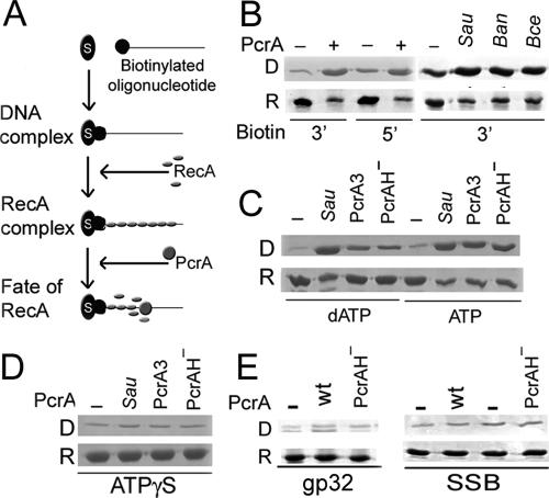
PcrA is a conserved DNA helicase present in all gram-positive bacteria. Bacteria lacking PcrA show high levels of recombination. Lethality induced by PcrA depletion can be overcome by suppressor mutations in the recombination genes recFOR. RecFOR proteins load RecA onto single-stranded DNA during recombination. Here we test whether an essential function of PcrA is to interfere with RecA-mediated DNA recombination in vitro. We demonstrate that PcrA can inhibit the RecA-mediated DNA strand exchange reaction in vitro. Furthermore, PcrA displaced RecA from RecA nucleoprotein filaments. Interestingly, helicase mutants of PcrA also displaced RecA from DNA and inhibited RecA-mediated DNA strand exchange. Employing a novel single-pair fluorescence resonance energy transfer-based assay, we demonstrate a lengthening of double-stranded DNA upon polymerization of RecA and show that PcrA and its helicase mutants can reverse this process. Our results show that the displacement of RecA from DNA by PcrA is not dependent on its translocase activity. Further, our results show that the helicase activity of PcrA, although not essential, might play a facilitatory role in the RecA displacement reaction.


Figures

FIG. 1.
FIG. 1.
Time dependence of RecA SE reaction and its inhibition by PcrA. (A) Schematic of RecA SE reaction. SSC DNA was coated with 6 μM RecA and mixed with DSL DNA. Transfer of the complementary ssDNA strand from DSL DNA to SSC DNA results in the formation of closed OC DNA and linear ssDNA. (B) Time course of SE reaction. RecA-mediated SE reaction was carried out for the indicated time periods, samples were run on a 1% agarose gel and transferred to a nylon membrane, and Southern blots were hybridized to labeled M13 RF DNA to visualize and quantify products. (C) Quantification of gel shown in panel B. Relative levels of DSL and OC DNA are shown as percentages of intensity of DSL DNA in the absence of RecA, which was taken to be 100%. Empty bars represent DSL DNA, while filled bars represent OC DNA. (D) Time dependence of inhibition of SE reaction by PcrA. Four hundred sixty nanomolar PcrA was added at various times after addition of DSL DNA. Samples were run on a 1% agarose gel containing ethidium bromide and visualized after extensive destaining. A negative image of the ethidium bromide-stained gel is shown. (E) Inhibition of SE reaction by PcrASau (Sau), PcrABan (Ban), or PcrABce (Bce) helicase. Increasing concentrations (4.6 nM, 46 nM, and 460 nM) of the various PcrA helicases were added to the SE reaction containing RecA-SSC DNA complex immediately before addition of dsDNA. A negative image of the gel is shown. (F) Quantification of SE inhibition by PcrASau, PcrABan, and PcrABce at the above three concentrations was carried out by transferring the DNA on the gel shown in panel E to a nylon membrane and hybridizing to labeled M13 RF DNA. Results are expressed as percent inhibition of the OC DNA product formed in the presence of various concentrations of different PcrA helicases. JM, joint molecule intermediates; OC, open-circular DNA; DSL, linear double-stranded DNA; SSC, circular ssDNA.
FIG. 2.
FIG. 2.
(A) Purification and characterization of His-PcrAH protein. (A) SDS-PAGE of purified His-PcrAH. M, molecular weight markers; U and I, protein lysates from uninduced or isopropyl-β-d-thiogalactopyranoside-induced cells, respectively; P, 500 ng purified His-PcrAH protein. (B) ATPase activity of PcrAH. Various concentrations of wild-type (wt) PcrA or PcrAH were tested for ATP hydrolysis in the presence or absence of ssDNA by TLC on polyethyleneimine-cellulose. (C) DNA binding activity of PcrAH. DNA binding activities of wild-type PcrA and PcrAH were analyzed by electrophoretic mobility shift assays using three different DNA probes. P, free probe; C, DNA-protein complex. (D) Helicase activities of PcrA and PcrAH in the presence of a folded DNA substrate. ds, partially double-stranded probe; ss, single-stranded product; C, DNA-protein complex. (E) Helicase activities of PcrA and PcrAH in the presence of partially duplex substrates containing either 3′ or 5′ ss dT tails. The structures of the various DNA substrates used are represented schematically at the bottom of the gel.
FIG. 3.
FIG. 3.
Inhibition of SE reaction by PcrA mutants. (A) Increasing concentrations (4.6 nM, 46 nM, and 460 nM) of PcrA or its mutants were added to the SE reaction and the products analyzed as described in the legend to Fig. 2A. (B) Quantification of SE inhibition by PcrA3Sau and PcrAH at the above three concentrations was carried out as described in the legend to Fig. 1E.
FIG. 4.
FIG. 4.
Displacement of RecA from ssDNA by PcrA. (A) Schematic of the assay. Bio-oligonucleotides were bound to streptavidin-coated magnetic beads. RecA was added to form the RecA-DNA-bead complex. PcrA was then added, and the fate of RecA was followed. (B) RecA displacement by PcrASau, PcrABan, and PcrABce. Row D indicates displaced RecA, and row R indicates RecA remaining on the beads. (C) RecA displacement by PcrASau from RecA-DNA complexes assembled in the presence of dATP or ATP. (D) Lack of RecA displacement by PcrASau when γ-S-ATP is substituted for ATP. (E) PcrASau does not displace the gp32 protein or SSB from ssDNA. wt, wild type.
FIG. 5.
FIG. 5.
PcrA displaces RecA from dsDNA. (A) Schematic of FRET-based assay for studying RecA displacement. Binding of RecA lengthens dsDNA 1.5-fold and decreases Eapp. Addition of PcrA displaces RecA from the DNA and increases the Eapp value. Distance 1× = 4.4 nm; 4n, four FRET pairs. (B to F) Histograms of Eapp of reactions as indicated. Means of Eapp values are indicated. Counts indicate numbers of individual DNA molecules. (G) PcrASau, PcrA3, and PcrAH display similar efficiencies of RecA displacement from dsDNA. The y axis represents the number of molecules displaying low Eapp values divided by that of those displaying high Eapp values.

References

    1. Aguilera, A. 2001. Double-strand break repair: are Rad51/RecA-DNA joints barriers to DNA replication? Trends Genet. 17:318-321. - PubMed
    1. Anand, S. P., A. Chattopadhyay, and S. A. Khan. 2005. The PcrA3 mutant binds DNA and interacts with the RepC initiator protein of plasmid pT181 but is defective in its DNA helicase and unwinding activities. Plasmid 54:104-113. - PubMed
    1. Anand, S. P., and S. A. Khan. 2004. Structure-specific DNA binding and bipolar helicase activities of PcrA. Nucleic Acids Res. 32:3190-3197. - PMC - PubMed
    1. Anand, S. P., P. Mitra, A. Naqvi, and S. A. Khan. 2004. Bacillus anthracis and Bacillus cereus PcrA helicases can support DNA unwinding and in vitro rolling-circle replication of plasmid pT181 of Staphylococcus aureus. J. Bacteriol. 186:2195-2199. - PMC - PubMed
    1. Bianco, P. R., and G. M. Weinstock. 1996. Interaction of the RecA protein of Escherichia coli with single-stranded oligodeoxyribonucleotides. Nucleic Acids Res. 24:4933-4939. - PMC - PubMed

Publication types

MeSH terms

LinkOut - more resources