Comparative Study
. 2007 Jun;189(12):4485-93.
doi: 10.1128/JB.01948-06. Epub 2007 Apr 20.

Cryo-electron tomography reveals the comparative three-dimensional architecture of Prochlorococcus, a globally important marine cyanobacterium

Affiliations
Comparative Study

Cryo-electron tomography reveals the comparative three-dimensional architecture of Prochlorococcus, a globally important marine cyanobacterium

Claire S Ting et al. J Bacteriol. 2007 Jun.

Abstract

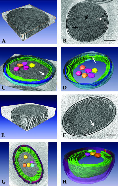
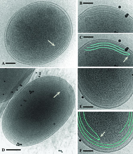
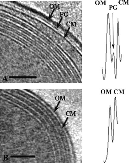
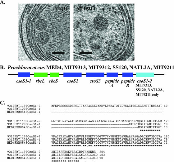
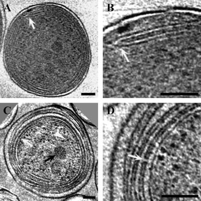
In an age of comparative microbial genomics, knowledge of the near-native architecture of microorganisms is essential for achieving an integrative understanding of physiology and function. We characterized and compared the three-dimensional architecture of the ecologically important cyanobacterium Prochlorococcus in a near-native state using cryo-electron tomography and found that closely related strains have diverged substantially in cellular organization and structure. By visualizing native, hydrated structures within cells, we discovered that the MED4 strain, which possesses one of the smallest genomes (1.66 Mbp) of any known photosynthetic organism, has evolved a comparatively streamlined cellular architecture. This strain possesses a smaller cell volume, an attenuated cell wall, and less extensive intracytoplasmic (photosynthetic) membrane system compared to the more deeply branched MIT9313 strain. Comparative genomic analyses indicate that differences have evolved in key structural genes, including those encoding enzymes involved in cell wall peptidoglycan biosynthesis. Although both strains possess carboxysomes that are polygonal and cluster in the central cytoplasm, the carboxysomes of MED4 are smaller. A streamlined cellular structure could be advantageous to microorganisms thriving in the low-nutrient conditions characteristic of large regions of the open ocean and thus have consequences for ecological niche differentiation. Through cryo-electron tomography we visualized, for the first time, the three-dimensional structure of the extensive network of photosynthetic lamellae within Prochlorococcus and the potential pathways for intracellular and intermembrane movement of molecules. Comparative information on the near-native structure of microorganisms is an important and necessary component of exploring microbial diversity and understanding its consequences for function and ecology.


Figures

FIG. 1.
FIG. 1.
Striking differences exist in the near-native cellular architecture of closely related Prochlorococcus strains. (A) 3D view of a single cell of Prochlorococcus MED4. (B) A 1.8-nm-thick tomographic slice of a MED4 cell. Two to three bands of intracytoplasmic (photosynthetic) lamellae are visible near the cell periphery (white arrow). Several polygonal structures resembling carboxysomes are clustered in the central cytoplasmic space (black arrows). (C) Surface-rendered model of a MED4 cell superimposed on a slice from a tomogram. Modeled 3D structures include the outer membrane (light blue), inner membrane (dark blue), intracytoplasmic lamellae (green), and carboxysomes (yellow, pink, orange). A distinct junction between two lamellae is visible (white arrow). (D) Surface-rendered model of MED4 showing that there are specific regions (white arrow) where the intracytoplasmic membranes (green) terminate, resulting in the formation of large gaps in the membrane bands. Note that an additional carboxysome is visible when the MED4 cell is viewed from this perspective. Color coding is as in panel C. (E) 3D view of a frozen-hydrated Prochlorococcus MIT9313 cell. (F) A 1.8-nm-thick tomographic slice of a MIT9313 cell. Several bands of intracytoplasmic lamellae are located near the cell membrane (white arrow). (G) Surface-rendered model of MIT9313 superimposed on a slice from a tomogram. Structures depicted include the cell wall (purple, pink, blue), extensive intracytoplasmic membrane system (green), and carboxysomes (yellow, pink, orange, blue). (H) Side view of a surface-rendered model of MIT9313, in which the regions where the intracytoplasmic membranes (green) terminate at the cell poles are visible. Note that these fenestrations in the intracytoplasmic membranes create areas of direct contact between the central cytoplasmic space and the region between the cell membrane and outermost intracytoplasmic membrane. Bars = 200 nm.
FIG. 2.
FIG. 2.
Prochlorococcus MED4 and MIT9313 cell structure as seen in frozen-hydrated whole mounts. In MED4, the intracytoplasmic membranes are typically apposed to one or two regions of the cell membrane (A, white arrow). (B and C) Higher magnifications of the internal membranes and a tracing (in blue) of their structure. A distinct junction between two intracytoplasmic membranes is shown (white arrow). (D, E, F) In MIT9313, the cell pole is the primary location for termination of the intracytoplasmic membranes and this results in prominent fenestrations (D, white arrow). (E) Structure of the intracytoplasmic membranes at a cell pole at a higher magnification; (F) tracing (in blue) of the same membranes. Note the distinct junction between two membranes (white arrow). In these images (B to F), the black particles are colloidal gold particles that were added to each sample and which serve as alignment fiducial markers. Bars = 100 nm (A, B, C, E, F) and 200 nm (D).
FIG. 3.
FIG. 3.
Prochlorococcus MED4 possesses a minimal cell wall architecture compared to MIT9313. Shown are 1.8-nm-thick slices from tomograms of frozen-hydrated MIT9313 (A) and MED4 (B) cells, with the cell wall area depicted. Cell wall structures indicated include the outer membrane (OM), peptidoglycan layer (PG), and cell membrane (CM). Note the layers of intracytoplasmic lamellae adjacent to the cell membrane in both cells. Density profiles of the wall region are shown adjacent to the corresponding cell images. The cell wall structure represented by a particular density peak is indicated, and these density profiles emphasize the differences in cell wall structure between MED4 and MIT9313. Bars = 100 nm.
FIG. 4.
FIG. 4.
The near-native structure of Prochlorococcus α-carboxysomes and the overall genomic organization of carboxysome shell polypeptide genes are highly conserved. (A) Carboxysomes of MED4 and MIT9313 are located typically in the central cytoplasmic space and are polygonal. Depicted are 1.8-nm-thick slices from a tomogram of a frozen-hydrated MED4 cell and a frozen-hydrated cryoultramicrotome section of a high-pressure-frozen MIT9313 cell. Bars = 100 nm. (B) The overall genomic organization of genes (csoS1-1, csoS2, csoS3, peptide A gene, peptide B gene) encoding putative carboxysome shell polypeptides is highly conserved in Prochlorococcus strains MED4, MIT9313, MIT9312, SS120, NATL2A, and MIT9211. Although the csoS1-2 gene is present in the genomes of strains (MIT9313, SS120, NATL2A, MIT9211) belonging to low-light-adapted Prochlorococcus clades, it is missing from the genomes of strains (MED4, MIT9312) that are members of high-light-adapted clades. rbcL and rbcS encode the large and small subunits of RuBisCO, respectively. (C) Alignment of the predicted amino acid sequence of CsoS1-1 from MED4 with CsoS1-1 and CsoS1-2 from MIT9313. Although these amino acid sequences are highly conserved (an asterisk indicates identical amino acid residues, a semicolon indicates conserved residues, and a period indicates semiconserved residues), CsoS1-2 possesses an extended N-terminal region.
FIG. 5.
FIG. 5.
Prochlorococcus intracytoplasmic lamellae are connected at distinct junctions and by membrane-lined channels. (A and B) Slices (1.8 nm thick) from a tomogram of a frozen-hydrated MED4 cell. The region in panel A at the white arrow is depicted at a higher magnification in panel B. Note the distinct junction between two intracytoplasmic membranes (B, white arrow). (C and D) Two different 1.8-nm tomographic slices of frozen-hydrated cryoultramicrotome sections of a high-pressure-frozen MIT9313 cell. The defocus (15 μm) and tilt angle (120°) were the same as those used for the frozen-hydrated whole-mount samples. Structures visible in panel C include carboxysomes (black arrow), ribosomes (white arrowhead), and tightly appressed intracytoplasmic lamellae (white arrow). Note that the luminal space of a lamella is less dense than the surrounding cytoplasm. A channel connecting adjacent intracytoplasmic lamellae is shown in panel D (white arrow). The apparent differences in the protein density of the cytoplasm between MED4 (A and B) and MIT9313 (C and D) reflect differences in contrast in the projection images of these samples. Comparisons of MED4 and MIT9313 cells that were prepared using the same technique of rapid freezing in liquid ethane followed by whole-mount imaging of frozen-hydrated samples indicate that the density of the cytoplasm is similar in these strains. Bars = 100 nm.

References

    1. Badger, M. R., and G. D. Price. 2003. CO2 concentrating mechanisms in cyanobacteria: molecular components, their diversity and evolution. J. Exp. Bot. 54:609-622. - PubMed
    1. Bergthorsson, U., and H. Ochman. 1998. Distribution of chromosome length variation in natural isolates of Escherichia coli. Mol. Biol. Evol. 15:9-16. - PubMed
    1. Campbell, L., H. B. Liu, H. A. Nolla, and D. Vaulot. 1997. Annual variability of phytoplankton and bacteria in the subtropical North Pacific Ocean at Station ALOHA during the 1991-1994 ENSO event. Deep-Sea Res. Part I 44:167.
    1. Crowther, R. A., D. J. DeRosier, and A. Klug. 1970. The reconstruction of a three-dimensional structure from its projections and its application to electron microscopy. Proc. R. Soc. Lond. A 317:319-340.
    1. Dmitriev, B., F. Toukach, and S. Ehlers. 2005. Towards a comprehensive view of the bacterial cell wall. Trends Microbiol. 13:569-574. - PubMed

Publication types

MeSH terms

LinkOut - more resources