. 2007 Jun;144(2):1012-28.
doi: 10.1104/pp.107.097477. Epub 2007 Apr 20.

A reevaluation of the key factors that influence tomato fruit softening and integrity

Affiliations

A reevaluation of the key factors that influence tomato fruit softening and integrity

Montserrat Saladié et al. Plant Physiol. 2007 Jun.

Abstract

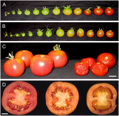
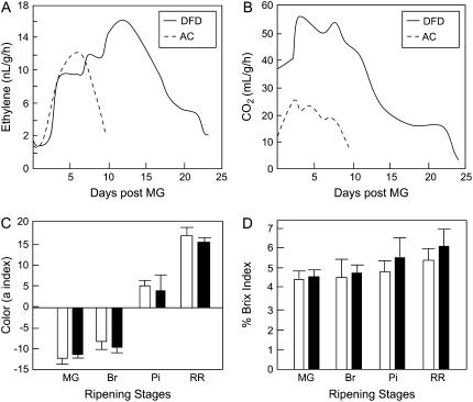
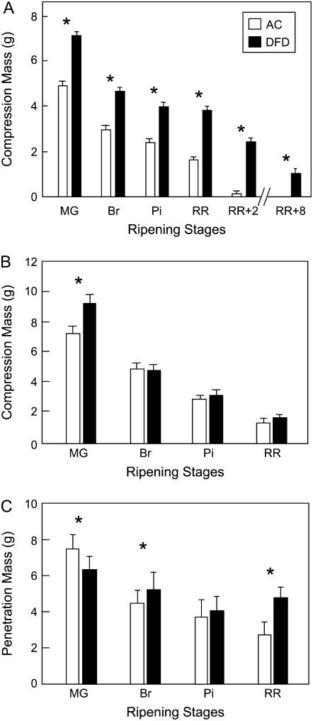
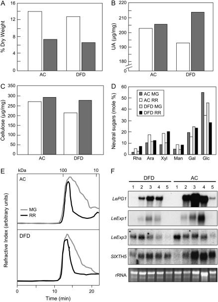
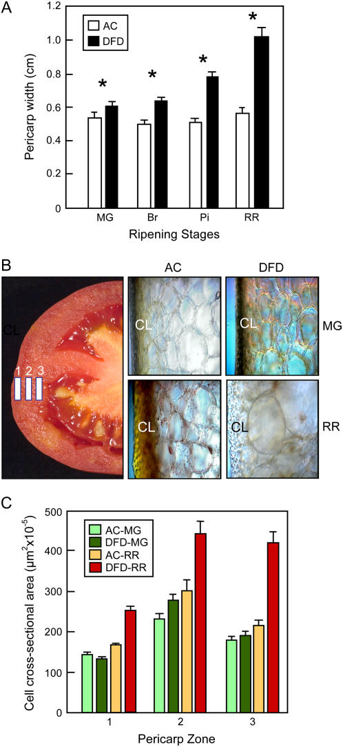
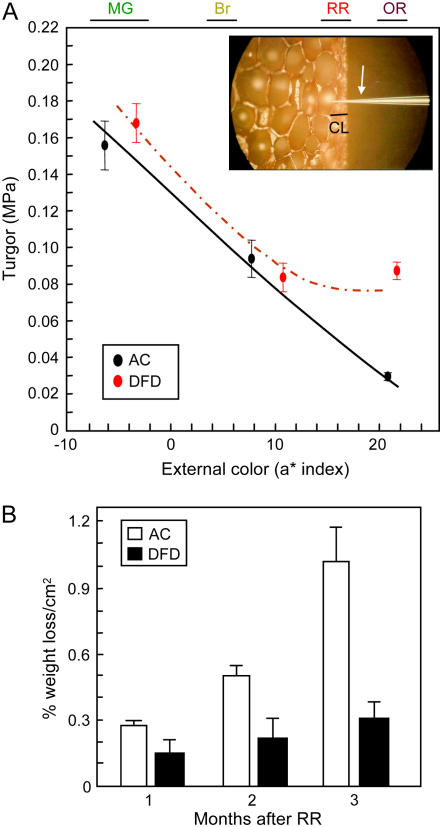
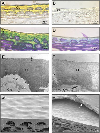
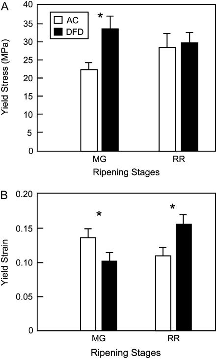
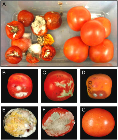
The softening of fleshy fruits, such as tomato (Solanum lycopersicum), during ripening is generally reported to result principally from disassembly of the primary cell wall and middle lamella. However, unsuccessful attempts to prolong fruit firmness by suppressing the expression of a range of wall-modifying proteins in transgenic tomato fruits do not support such a simple model. 'Delayed Fruit Deterioration' (DFD) is a previously unreported tomato cultivar that provides a unique opportunity to assess the contribution of wall metabolism to fruit firmness, since DFD fruits exhibit minimal softening but undergo otherwise normal ripening, unlike all known nonsoftening tomato mutants reported to date. Wall disassembly, reduced intercellular adhesion, and the expression of genes associated with wall degradation were similar in DFD fruit and those of the normally softening 'Ailsa Craig'. However, ripening DFD fruit showed minimal transpirational water loss and substantially elevated cellular turgor. This allowed an evaluation of the relative contribution and timing of wall disassembly and water loss to fruit softening, which suggested that both processes have a critical influence. Biochemical and biomechanical analyses identified several unusual features of DFD cuticles and the data indicate that, as with wall metabolism, changes in cuticle composition and architecture are an integral and regulated part of the ripening program. A model is proposed in which the cuticle affects the softening of intact tomato fruit both directly, by providing a physical support, and indirectly, by regulating water status.


Figures

Figure 1.
Figure 1.
Comparison of AC and DFD fruit phenotypes. Developmental series of AC fruits (A) and DFD fruits (B). C, DFD (left group) and AC (right group) fruits after 4 months of storage at room temperature. Scale bar = 2 cm. D, DFD tomatoes bisected after 3 (left), 6 (middle), and 7 (right) months of storage at room temperature. Scale bar = 1 cm.
Figure 2.
Figure 2.
Ripening-related traits in AC and DFD fruits. Ethylene (A) and carbon dioxide (B) production by ripening AC and DFD fruits detached at the MG stage. C, Evaluation of external ripening-related color change of AC and DFD fruits (for C and D, mean ± se, n = 20). D, Total soluble solids in the pericarp of AC (black bars) and DFD (white bars) fruits at the MG, Br, Pi, and RR stages, measured using a refractometer.
Figure 3.
Figure 3.
Textural analysis of tomato fruits. Compression test of intact tomato fruit (A) and excised pericarp segments (B). C, Penetration analysis of combined pericarp and cuticle. Fruits and pericarp segments from AC and DFD were tested at MG, Br, Pi, and RR stages and at RR plus 2 or 8 months (RR + 2 or RR + 8, respectively). Asterisks indicate statistically significance differences (mean ± se, n = 20, P < 0.001).
Figure 4.
Figure 4.
Cell wall analysis and wall metabolism-related gene expression. Relative amounts of cell wall pellet (A), uronic acid (UA; wall dry weight basis) content (B), cellulose content (C), and neutral sugar composition (D; mole percent of total wall noncellulosic neutral sugars) of isolated pericarp from AC and DFD fruits at the MG (white bars) and RR (gray bars) stages. For D, individual sugars comprised Rha, Ara, Xyl, Man, Gal, and Glc. E, SEC of water-soluble pectins from AC and DFD fruits at the MG and RR stages. Molecular mass calibration markers (kilodaltons) are shown at the top. F, RNA gel-blot analysis of PG (LePG1), expansins (LeExp1, LeExp3), and xyloglucan endotransglucosylase/hydrolase (SlXTH5) expression in DFD and AC pericarp. Lanes 1 to 5 correspond to the ripening stages MG, Br, Pi, RR, and OR, respectively. Ethidium bromide-stained rRNA in the same gel is shown as a loading control.
Figure 5.
Figure 5.
Pericarp and cell size measurements. A, Pericarp width of AC and DFD fruits at the MG, Br, Pi, and RR stages. Asterisks indicate statistically significance differences (mean ± se, n = 20). B, Bisected RR tomato illustrating the three pericarp zones in which cell size was measured. The adjacent light microscopy photographs show cells in zone 1 and the cuticular layer (CL) from DFD and AC fruits at the MG and RR stages. C, Cell cross-sectional area in the three pericarp zones.
Figure 6.
Figure 6.
Pericarp cellular turgor pressure evaluation and fruit water loss. A, Turgor pressure of outer pericarp cells from AC and DFD fruits at different ripening stages, as determined by external color (a* index value). The color ranges of MG, Br, RR, and OR fruit are shown. A smoothing spline regression curve and the mean and se (n3 5) is shown for each cultivar. A photograph of the microprobe (white arrow) penetrating the cuticular layer (CL) and an outer pericarp cell is shown in the inset. B, Percentage water loss from detached AC and DFD fruits over a 3 month period after reaching the RR stage (mean ± se, n = 10).
Figure 7.
Figure 7.
Microscopy analyses of AC and DFD fruit cuticles. Light microscopy images without (A and B) or with (C and D) toluidine blue O staining and TEM (E and F) and SEM (G and H) images of AC (A, C, E, and G) and DFD (B, D, F, and H) RR stage cuticles and outer pericarp. CL, Cuticular layer; CW, cell wall; cytosol, Cyt. The arrow in H indicates a thin membranous layer on the DFD fruit cuticle surface. Scale bars for A to D = 5 μm; E and F = 0.5 μm; G = 10 μm; and H = 50 μm.
Figure 8.
Figure 8.
Instron analysis of isolated tomato fruit cuticles. The yield stress (A) and yield strain (B) of cuticles from AC and DFD fruit at the MG and RR stages, evaluated with an Instron testing machine (asterisks indicate statistically significance differences within a ripening stage; mean ± se, n = 20).
Figure 9.
Figure 9.
Microbial infection of AC and DFD fruits. A, RR fruits from AC (left side) and DFD (right side) stored for 5 months in high humidity and temperature conditions. Ripe fruits from a commercial cultivar (B and E), AC (C and F), and DFD (D and G) following inoculation with three concentrations of B. cinerea spores through surface penetrations (B–D) or applied ectopically (E–G).

References

    1. Ahmed AER, Labavitch JM (1977) A simplified method for accurate determination of cell wall uronic acid content. J Food Biochem 1 361–365
    1. Almeida DPF, Huber DJ (1999) Apoplastic pH and inorganic ion levels in tomato fruit: a potential means for regulation of cell wall metabolism during ripening. Physiol Plant 105 506–512
    1. Baker EA, Bukovac MJ, Hunt GM (1982) Composition of tomato fruit cuticle as related to fruit growth and development. In DF Cutler, KL Alvin, CE Price, eds, The Plant Cuticle. Academic Press, London, pp 33–44
    1. Banik M, Bourgault R, Bewley JD (2001) Endo-β-mannanase is present in an inactive form in ripening tomato fruits of the cultivar Walter. J Exp Bot 52 105–111 - PubMed
    1. Bargel H, Neinhuis C (2004) Altered tomato (Lycopersicon esculentum Mill.) fruit cuticle biomechanics of a pleiotropic non ripening mutant. J Plant Growth Regul 23 61–75

Publication types

MeSH terms

LinkOut - more resources