

Water transport in aquaporins: osmotic permeability matrix analysis of molecular dynamics simulations
- PMID: 17449664
- PMCID: PMC1896254
- DOI: 10.1529/biophysj.106.101170
Water transport in aquaporins: osmotic permeability matrix analysis of molecular dynamics simulations
Abstract
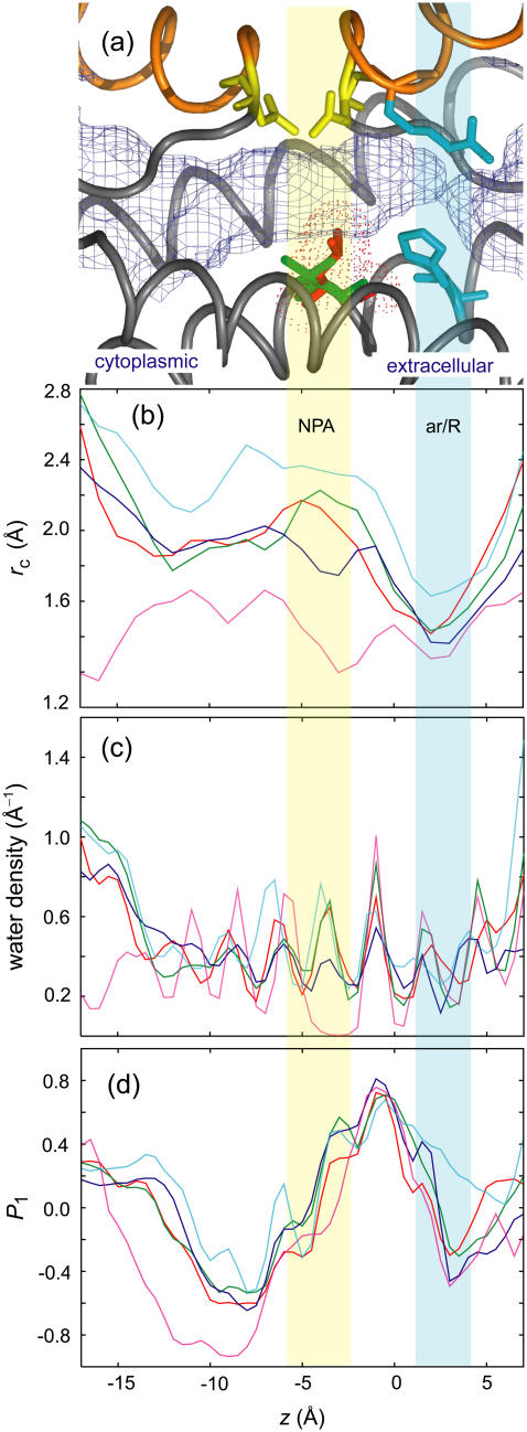
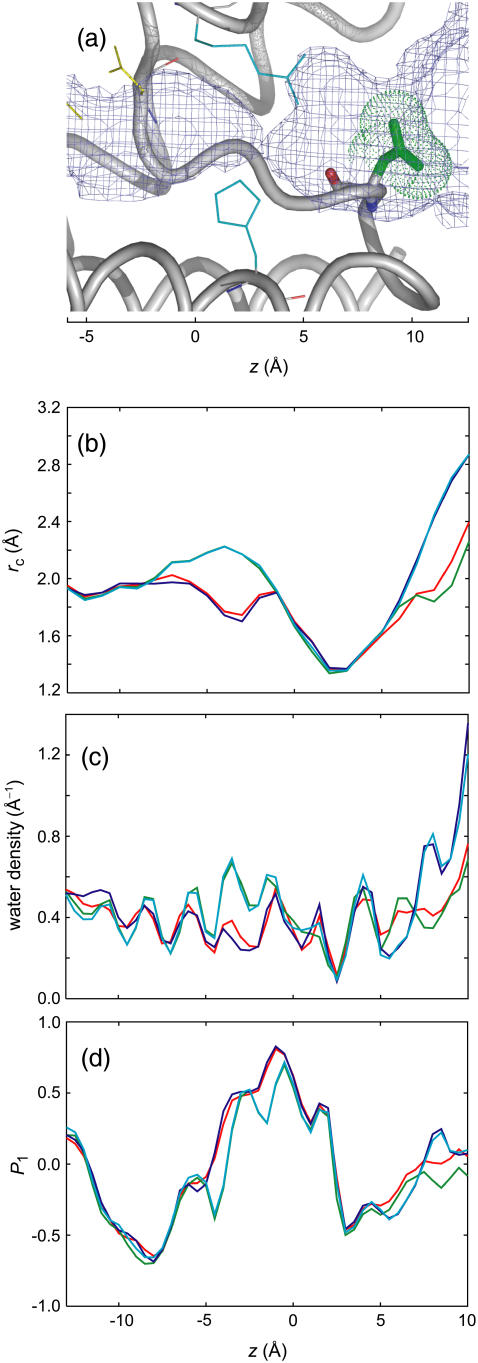
Single-channel osmotic water permeability (p(f)) is a key quantity for investigating the transport capability of the water channel protein, aquaporin. However, the direct connection between the single scalar quantity p(f) and the channel structure remains unclear. In this study, based on molecular dynamics simulations, we propose a p(f)-matrix method, in which p(f) is decomposed into contributions from each local region of the channel. Diagonal elements of the p(f) matrix are equivalent to the local permeability at each region of the channel, and off-diagonal elements represent correlated motions of water molecules in different regions. Averaging both diagonal and off-diagonal elements of the p(f) matrix recovers p(f) for the entire channel; this implies that correlated motions between distantly-separated water molecules, as well as adjacent water molecules, influence the osmotic permeability. The p(f) matrices from molecular dynamics simulations of five aquaporins (AQP0, AQP1, AQP4, AqpZ, and GlpF) indicated that the reduction in the water correlation across the Asn-Pro-Ala region, and the small local permeability around the ar/R region, characterize the transport efficiency of water. These structural determinants in water permeation were confirmed in molecular dynamics simulations of three mutants of AqpZ, which mimic AQP1.
Figures


Similar articles
-
Comparative simulations of aquaporin family: AQP1, AQPZ, AQP0 and GlpF.FEBS Lett. 2005 Oct 24;579(25):5549-52. doi: 10.1016/j.febslet.2005.09.018. FEBS Lett. 2005. PMID: 16225876
-
Single-channel water permeabilities of Escherichia coli aquaporins AqpZ and GlpF.Biophys J. 2006 Apr 1;90(7):2270-84. doi: 10.1529/biophysj.105.073965. Epub 2006 Jan 6. Biophys J. 2006. PMID: 16399837 Free PMC article.
-
Water transport in aquaporins: molecular dynamics simulations.Front Biosci (Landmark Ed). 2009 Jan 1;14(4):1283-91. doi: 10.2741/3308. Front Biosci (Landmark Ed). 2009. PMID: 19273130 Review.
-
Dynamic and energetic mechanisms for the distinct permeation rate in AQP1 and AQP0.Biochim Biophys Acta. 2010 Mar;1798(3):318-26. doi: 10.1016/j.bbamem.2009.11.015. Epub 2009 Dec 2. Biochim Biophys Acta. 2010. PMID: 19961829
-
Dynamics and energetics of permeation through aquaporins. What do we learn from molecular dynamics simulations?Handb Exp Pharmacol. 2009;(190):57-76. doi: 10.1007/978-3-540-79885-9_3. Handb Exp Pharmacol. 2009. PMID: 19096772 Review.
Cited by
-
Capturing Functional Motions of Membrane Channels and Transporters with Molecular Dynamics Simulation.J Comput Theor Nanosci. 2010 Dec;7(12):2481-2500. doi: 10.1166/jctn.2010.1636. J Comput Theor Nanosci. 2010. PMID: 23710155 Free PMC article.
-
Optimizing water permeability through the hourglass shape of aquaporins.Proc Natl Acad Sci U S A. 2013 Oct 8;110(41):16367-72. doi: 10.1073/pnas.1306447110. Epub 2013 Sep 25. Proc Natl Acad Sci U S A. 2013. PMID: 24067650 Free PMC article.
-
Mechanism of the αβ conformational change in F1-ATPase after ATP hydrolysis: free-energy simulations.Biophys J. 2015 Jan 6;108(1):85-97. doi: 10.1016/j.bpj.2014.11.1853. Biophys J. 2015. PMID: 25564855 Free PMC article.
-
Water in Nanopores and Biological Channels: A Molecular Simulation Perspective.Chem Rev. 2020 Sep 23;120(18):10298-10335. doi: 10.1021/acs.chemrev.9b00830. Epub 2020 Aug 25. Chem Rev. 2020. PMID: 32841020 Free PMC article. Review.
-
Statistical thermodynamics of biomembranes.Cryobiology. 2010 Feb;60(1):80-90. doi: 10.1016/j.cryobiol.2009.05.001. Epub 2009 May 19. Cryobiology. 2010. PMID: 19460363 Free PMC article.
References
-
- Borgnia, M., S. Nielsen, A. Engel, and P. Agre. 1999. Cellular and molecular biology of the aquaporin water channels. Annu. Rev. Biochem. 68:425–458. - PubMed
-
- Murata, K., K. Mitsuoka, T. Hirai, T. Walz, P. Agre, J. B. Heymann, A. Engel, and Y. Fujiyoshi. 2000. Structural determinants of water permeation through aquaporin-1. Nature. 407:599–605. - PubMed
-
- Fu, D., A. Libson, L. J. Miercke, C. Weitzman, P. Nollert, J. Krucinski, and R. M. Stroud. 2000. Structure of a glycerol-conducting channel and the basis for its selectivity. Science. 290:481–486. - PubMed
-
- Sui, H., B. G. Han, J. K. Lee, P. Walian, and B. K. Jap. 2001. Structural basis of water-specific transport through the AQP1 water channel. Nature. 414:872–878. - PubMed
Publication types
MeSH terms
Substances
LinkOut - more resources
Full Text Sources
Research Materials
