Comparative Study
. 2007 Jun;73(11):3695-704.
doi: 10.1128/AEM.02735-06. Epub 2007 Apr 20.

Detection and transformation of genome segments that differ within a coastal population of Vibrio cholerae strains

Affiliations
Comparative Study

Detection and transformation of genome segments that differ within a coastal population of Vibrio cholerae strains

Michael C Miller et al. Appl Environ Microbiol. 2007 Jun.

Abstract

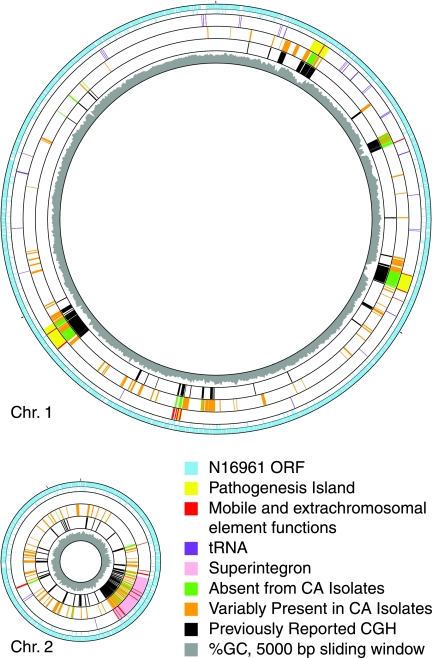
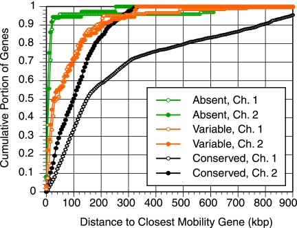
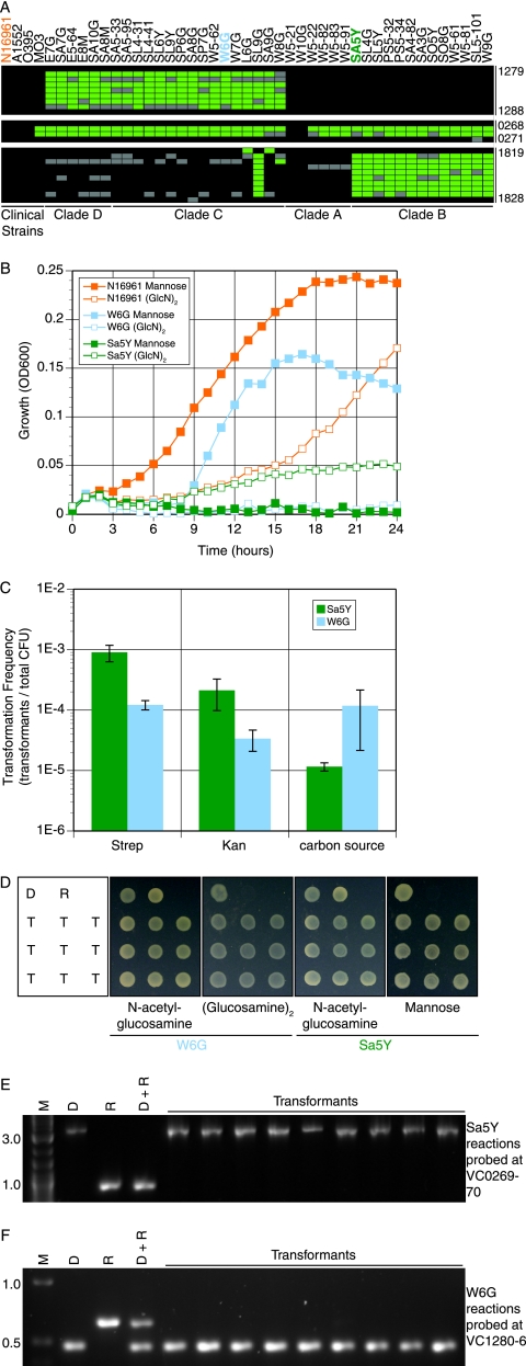
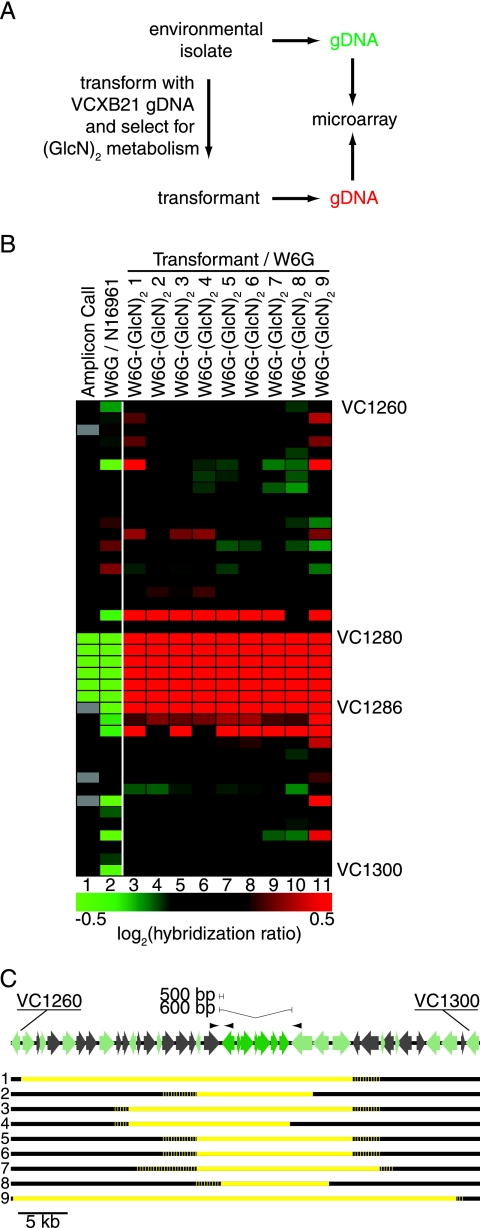
Vibrio cholerae is an autochthonous member of diverse aquatic ecosystems around the globe. Collectively, the genomes of environmental V. cholerae strains comprise a large repository of encoded functions which can be acquired by individual V. cholerae lineages through uptake and recombination. To characterize the genomic diversity of environmental V. cholerae, we used comparative genome hybridization to study 41 environmental strains isolated from diverse habitats along the central California coast, a region free of endemic cholera. These data were used to classify genes of the epidemic V. cholerae O1 sequenced strain N16961 as conserved, variably present, or absent from the isolates. For the most part, absent genes were restricted to large mobile elements and have known functions in pathogenesis. Conversely, genes present in some, but not all, California isolates were in smaller contiguous clusters and were less likely to be near genes with functions in DNA mobility. Two such clusters of variable genes encoding different selectable metabolic phenotypes (mannose and diglucosamine utilization) were transformed into the genomes of environmental isolates by chitin-dependent competence, indicating that this mechanism of general genetic exchange is conserved among V. cholerae. The transformed DNA had an average size of 22.7 kbp, demonstrating that natural competence can mediate the movement of large chromosome fragments. Thus, whether variable genes arise through the acquisition of new sequences by horizontal gene transfer or by the loss of preexisting DNA though deletion, natural transformation provides a mechanism by which V. cholerae clones can gain access to the V. cholerae pan-genome.


Figures

FIG. 1.
FIG. 1.
Genes variably present in or absent from all California Vibrio cholerae isolates mapped onto N16961 chromosomes. Circle one shows each of the protein-coding genes (16) in reference strain N16961 probed in this study (blue); circle two shows genes not probed. Circle three shows genome landmarks. Absent (green) and variable (orange) genes are plotted in the fourth circle. Conserved and uncalled genes are not shown. Circle five shows genes identified by previous CGH studies as missing from at least one V. cholerae isolate (black) (8, 9). Circle six shows the percentage of G+C (gray) in a 5,000-bp window in 1,000-bp steps, determined using GACK software (28). Generated with GenoMap (47).
FIG. 2.
FIG. 2.
Distance to mobility genes. The distance from the midpoint of each gene probed by CGH to the midpoint of the closest N16961 gene annotated as “mobile and extrachromosomal element functions” (16) is plotted in a cumulative histogram. Absent (green), variable (orange), and conserved (black) genes are plotted by chromosome. Because the chromosomes vary in size, the maximum possible distances are 1,481 kbp for chromosome 1 (Ch. 1) and 536 kbp for Ch. 2. This, along with uneven distribution of mobility genes, accounts for much of the differences between the shapes of the curves for the conserved genes.
FIG. 3.
FIG. 3.
Transformation of variable loci. (A) Positive (black), negative (green), or uncertain (gray) genes at three loci on chromosome 1 for each isolate genome. Strains are grouped by overall genomic similarity based on a report by Keymer et al. (27). (B) Growth in M9 with 0.001% amino acids and 0.2% mannose (filled symbols) or (GlcN)2 (open symbols). W6G (blue) lacks genes VC1280 to VC1286. Sa5Y (green) lacks both VC0269 to VC0270 and VC01820 to VC01827. N16961 (orange) encodes all three loci. (C) Isolates Sa5Y (green) and W6G (blue) were grown on crab shell and transformed with gDNA from strain VCXB21. Transformants were selected on LB medium containing the indicated antibiotic or M9 with (GlcN)2 (W6G) or mannose (Sa5Y). (D) Metabolic phenotypes of transformants were confirmed by growth on M9 containing the indicated carbon source. D, gDNA donor strain; R, recipient isolate; T, nine independent transformants. (E and F) The selected locus transformed from the donor strain into the recipient isolate was detected by PCR. M, molecular weight marker (kbp). (E) Probes VC0269 to VC0270 transformed into Sa5Y using primers in VC0268, an intervening gene present in Sa5Y but not in VCXB21, and VC0271. (F) Probes VC1280 to VC1286 transformed into W6G using primers in VC1279, VC1280, and VC1287.
FIG. 4.
FIG. 4.
Recombination of large chromosome fragments at VC1260 to VC1300 by naturally competent V. cholerae. (A) Strategy for detecting transformed DNA by microarray hybridization. (B) Oligonucleotide microarray CGH comparison of W6G transformants selected for growth on (GlcN)2 to the original W6G isolate. Lane 1 is colored as in Fig. 3A and reflects processed calls, not raw hybridization data. Lane 2 is colored to reflect W6G (red) hybridization relative to N16961 (green). Lanes 3 to 11 reflect transformant hybridization (red) relative to that of environmental isolate W6G (green) as depicted in panel A. (C) Dark green ORFs (VC1280 to VC1286) are missing from W6G and were acquired by the transformants. Light green ORFs are present in W6G but can be distinguished from homologous genes in VCXB21 by virtue of reduced microarray hybridization and/or sequenced single-nucleotide polymorphisms. For each of nine transformants, yellow reflects DNA donated from VCXB21, and black reflects DNA of W6G origin. Recombination junctions were resolved to the gene. The dotted line reflects DNA of ambiguous origin due to a lack of sequenced single-nucleotide polymorphisms. The arrowheads above ORF map reflect primers used in Fig. 3F.

References

    1. Ansaruzzaman, M., M. J. Albert, I. Kuhn, S. M. Faruque, A. K. Siddique, and R. Mollby. 1996. Differentiation of Vibrio cholerae O1 isolates with biochemical fingerprinting and comparison with ribotyping. J. Diarrhoeal Dis. Res. 14:248-254. - PubMed
    1. Ausubel, F., R. Brent, R. E. Kingston, D. D. Moore, J. G. Seidman, J. A. Smith, and K. Struhl. 1997. Short protocols in molecular biology. Wiley, New York, NY.
    1. Beltran, P., G. Delgado, A. Navarro, F. Trujillo, R. K. Selander, and A. Cravioto. 1999. Genetic diversity and population structure of Vibrio cholerae. J. Clin. Microbiol. 37:581-590. - PMC - PubMed
    1. Bik, E. M., R. D. Gouw, and F. R. Mooi. 1996. DNA fingerprinting of Vibrio cholerae strains with a novel insertion sequence element: a tool to identify epidemic strains. J. Clin. Microbiol. 34:1453-1461. - PMC - PubMed
    1. Byun, R., L. D. H. Elbourne, R. Lan, and P. R. Reeves. 1999. Evolutionary relationships of pathogenic clones of Vibrio cholerae by sequence analysis of four housekeeping genes. Infect. Immun. 67:1116-1124. - PMC - PubMed

Publication types

MeSH terms