. 2007 Apr;19(4):1221-34.
doi: 10.1105/tpc.106.048264. Epub 2007 Apr 20.

An ERF transcription factor in Medicago truncatula that is essential for Nod factor signal transduction

Affiliations

An ERF transcription factor in Medicago truncatula that is essential for Nod factor signal transduction

Patrick H Middleton et al. Plant Cell. 2007 Apr.

Abstract

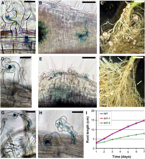
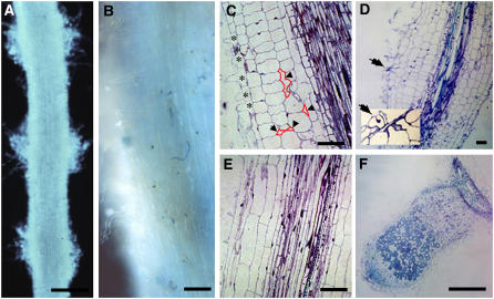
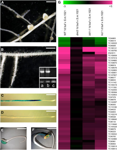
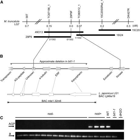
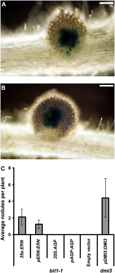
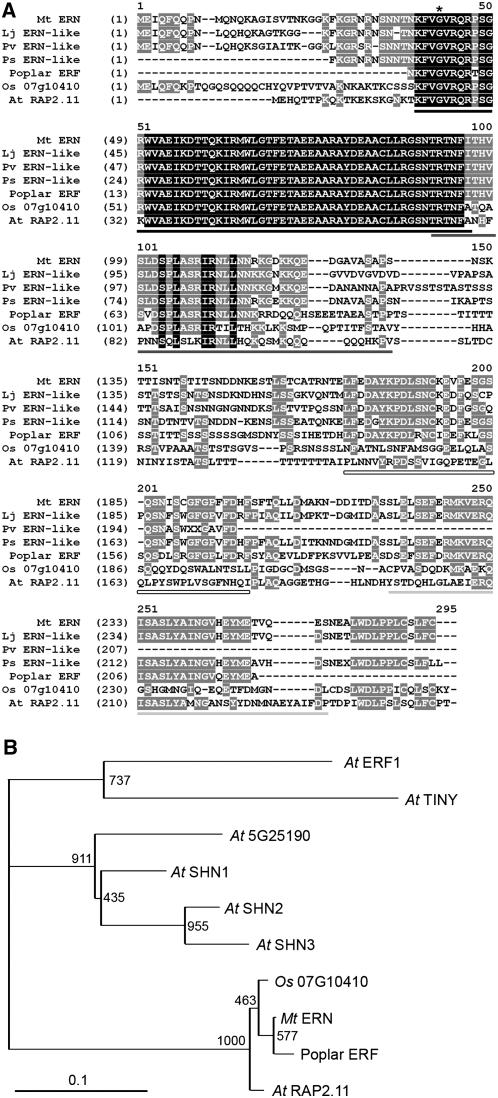
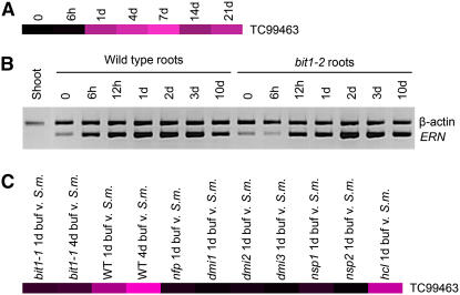
Rhizobial bacteria activate the formation of nodules on the appropriate host legume plant, and this requires the bacterial signaling molecule Nod factor. Perception of Nod factor in the plant leads to the activation of a number of rhizobial-induced genes. Putative transcriptional regulators in the GRAS family are known to function in Nod factor signaling, but these proteins have not been shown to be capable of direct DNA binding. Here, we identify an ERF transcription factor, ERF Required for Nodulation (ERN), which contains a highly conserved AP2 DNA binding domain, that is necessary for nodulation. Mutations in this gene block the initiation and development of rhizobial invasion structures, termed infection threads, and thus block nodule invasion by the bacteria. We show that ERN is necessary for Nod factor-induced gene expression and for spontaneous nodulation activated by the calcium- and calmodulin-dependent protein kinase, DMI3, which is a component of the Nod factor signaling pathway. We propose that ERN is a component of the Nod factor signal transduction pathway and functions downstream of DMI3 to activate nodulation gene expression.


Figures

Figure 1.
Figure 1.
bit1-1 Shows Defects in Infection Thread Initiation and Maintenance. (A) to (F) S. meliloti infection in wild-type ([A] to [C]) and bit1-1 ([D] to [F]) plants. (A) Five days after inoculation with S. meliloti, the infection threads on wild-type plants have exited the root hair cell and begin to divide toward the cortex. (B) One week after inoculation, the wild-type infection threads have ramified into the dividing cells of the nodule primordium. (C) After 2 months in the soil with S. meliloti, large pink nodules can be seen on wild-type plants. (D) By contrast, bit1-1 mutants show only infection foci development within the first week of infection with S. meliloti. (E) Three weeks after infection, bit1-1 mutants show some cortical cell division, and infection threads may invade the first cortical cell layer. (F) After 2 months in the soil with S. meliloti, bit1-1 plants show small undeveloped nodules, indicated with arrows. (G) and (H) Infection threads that do initiate on bit1-1 are often branched. (I) The root growth of bit1-2 (green) is much slower than either bit1-1 (red) or the wild type (blue). Bars represent se. Bacteria were visualized using X-galactosidase staining ([A], [B], [D], [E], [G], and [H]). Bars = 20 μm in (A), (D), (G), and (H), 100 μm in (B) and (E), and 5 mm in (C) and (F).
Figure 2.
Figure 2.
Cytological Analysis Reveals bit1-2 Phenotypes Affecting Both Nodulation and Root Development. Light micrographs of roots of bit1-2 ([A] to [D]) and the wild type ([E] and [F]). (A), (C), and (E) are noninoculated roots. (B) is 3 d after inoculation, and (D) and (F) are 21 d after inoculation. In (B), numerous infections can be observed at 3 d after inoculation, although these infections uniformly arrest within the epidermis. The inset in (D) shows a higher magnification of an arrested infection (arrow) at 21 d after inoculation. Note that in addition to altered cortical cell dimensions, bit1-2 is characterized by close spacing of root hairs ([C]; stars) and the presence of intercellular spaces between cortical cells ([C]; arrowheads with selected intracellular spaces outlined in red for clarity). Radial root swelling shown for bit1-2 at 21 d after inoculation was correlated with altered cortical cell morphology but not S. meliloti–induced cell division. Bars = 500 μm in (A) and (F) and 100 μm in (B) to (E).
Figure 3.
Figure 3.
bit1 Is Defective in Nod Factor Signaling. (A) and (B) Spontaneous nodules form in the absence of S. meliloti in plants transformed with the constitutively active kinase of DMI3, DMI31-311 (A), but not in bit1-1 plants transformed with DMI31-311 (B), indicating that BIT1 is necessary for DMI31-311-induced spontaneous nodulation. To ensure that bit1 plants were transformed with DMI31-311, we assessed for the presence of the kanamycin gene in transformed root systems ([B], top panel of inset): (a) bit1-1, (b) bit1-2, and (c) wild type; the bottom panel shows amplification of a positive control. Bars = 5 mm. (C) and (D) Treatment with 1 nM Nod factor leads to the induction of ENOD11:GUS in wild-type (C) but not bit1-1 (D) plants. (E) and (F) Nonsymbiotic ENOD11:GUS activity is present in the cotyledons of both wild-type (E) and bit1-1 (F) plants. Bars = 10 mm in (C) to (F). (G) Forty-six tentative consensus sequences (TC) previously identified as being induced/repressed by Nod factor and S. meliloti within 24 h of treatment were assessed in bit1-1 using Affymetrix GeneChips. The induction/repression of these tentative consensus sequences is shown in the wild type, dmi3-1, bit1-1, and hcl (the data for dmi3 and hcl have been reported previously in Mitra et al., 2004a). A pooled log2 fold change was calculated from three replicates of buffer-treated versus S. meliloti 1021–treated plants. The brightest green represents a log2 fold change of −3, and the brightest magenta a log2 fold change of +3. Analysis was performed using SAM2.11; for q-values of data, see Supplemental Table 1 online.
Figure 4.
Figure 4.
BIT1 Identification by Positional and Transcript-Based Cloning. (A) and (B) The bit1-2 mutation was found to be located between the genetic markers TC87156_1 and TC80512_1 on linkage group 7 (A). The physical region between these markers is spanned by the three BACs 29P9, 49O13, and 16I24 and contains four genes (B). The deletion that causes the bit1-1 mutation covers a similar region as indicated. A region on L. japonicus linkage group 1 shows absolute colinearity. As shown, the bit1-1 deletion affects at least five genes. (C) Amplification of a 200-bp fragment of TC102418 encoding the AGP contained within the bit1-1 deletion in a segregating F2 backcrossed population of bit1-1 reveals cosegregation of the deletion with the nod phenotype. Bottom panel (a) shows amplification of a positive control.
Figure 5.
Figure 5.
Complementation of the bit1-1 Phenotype with ERN. (A) and (B) The transformation of bit1-1 plants with pERN:ERN (ERN driven by its native promoter) leads to complementation of the mutant allowing nodulation after S. meliloti infection. X-galactosidase staining reveals S. meliloti contained within infection threads that ramify throughout the nodule primordium. Bars = 50 μm. (C) Average number of nodules on all transformed plants, including those showing no nodulation, 20 d after inoculation with S. meliloti. bit1-1 plants were transformed with five constructs; the ERN gene regulated by the 35S promoter or its native promoter and the AGP that is also contained within the bit1-1 deletion regulated by the 35S promoter or its native promoter and an empty vector. As a positive control, dmi3 plants were complemented with DMI3 driven by its native promoter. Error bars represent se. There is no significant difference (P = 0.1) among 35S:ERN, pERN:ERN, and pDMI3:DMI3.
Figure 6.
Figure 6.
M. truncatula ERN Is an ERF Transcription Factor. (A) Alignments of M. truncatula ERN with homologs form L. japonicus, P. sativum, P. vulgaris, poplar, rice, and Arabidopsis. The AP2 domain is underlined in black, CMV-3 is underlined in dark gray, and CMV-4 is underlined in light gray. A novel conserved domain that is specific to plants in the Rosid 1 clade is also apparent and underlined by a hollow bar. The asterisk denotes the site of the bit1-2 mutation. (B) A phylogenetic tree of the AP2 domains of M. truncatula ERN and its homologs. Since the AP2 domains of the legume homologs are identical, only M. truncatula ERN is shown along with the AP2 domains of rice, poplar, the group V Arabidopsis ERFs, and the Arabidopsis ERFs TINY and ERF1 as outgroups.
Figure 7.
Figure 7.
ERN Is Induced upon Rhizobial Inoculation, and This Requires the Nod Factor Signaling Pathway. (A) Affymetrix GeneChip analysis reveals that TC99463, which encodes ERN, is induced following S. meliloti inoculation, with maximal induction at 7 d after infection. Expression levels were calculated by comparing three replicates of infected plants with three replicates of uninfected plants. Black represents no induction and the brightest magenta a 2.0 log2 fold change. For q-values, see Supplemental Table 3 online. (B) RT-PCR of ERN reveals induction following S. meliloti inoculation, with maximal induction at 72 h in the time points assessed in both wild-type and bit1-2 roots. (C) Affymetrix analysis of the induction of ERN (TC99463) at 24 h after rhizobial infection requires components of the Nod factor signaling pathway but not HCL. Black represents no induction and the brightest magenta a 1.7 log2 fold change compared with uninfected plants. For q-values, see Supplemental Table 3 online. Some data from (A) and (C) have been published previously (Mitra et al., 2004a; Starker et al., 2006).

References

    1. Aharoni, A., Dixit, S., Jetter, R., Thoenes, E., van Arkel, G., and Pereira, A. (2004). The SHINE clade of AP2 domain transcription factors activates wax biosynthesis, alters cuticle properties, and confers drought tolerance when overexpressed in Arabidopsis. Plant Cell 16 2463–2480. - PMC - PubMed
    1. Allen, M.D., Yamasaki, K., Ohme-Takagi, M., Tateno, M., and Suzuki, M. (1998). A novel mode of DNA recognition by a beta-sheet revealed by the solution structure of the GCC-box binding domain in complex with DNA. EMBO J. 17 5484–5496. - PMC - PubMed
    1. Ane, J.M., et al. (2004). Medicago truncatula DMI1 required for bacterial and fungal symbioses in legumes. Science 303 1364–1367. - PubMed
    1. Ardourel, M., Demont, N., Debelle, F.D., Maillet, F., Debilly, F., Prome, J.C., Denarie, J., and Truchet, G. (1994). Rhizobium meliloti lipooligosaccharide nodulation factors: Different structural requirements for bacterial entry into target root hair cells and induction of plant symbiotic developmental responses. Plant Cell 6 1357–1374. - PMC - PubMed
    1. Arrighi, J.F., et al. (2006). The Medicago truncatula lysine motif-receptor-like kinase gene family includes NFP and new nodule-expressed genes. Plant Physiol. 142 265–279. - PMC - PubMed

Publication types

MeSH terms