. 2007 Apr;19(4):1415-31.
doi: 10.1105/tpc.106.042291. Epub 2007 Apr 20.

SCABP8/CBL10, a putative calcium sensor, interacts with the protein kinase SOS2 to protect Arabidopsis shoots from salt stress

Affiliations

SCABP8/CBL10, a putative calcium sensor, interacts with the protein kinase SOS2 to protect Arabidopsis shoots from salt stress

Ruidang Quan et al. Plant Cell. 2007 Apr.

Abstract

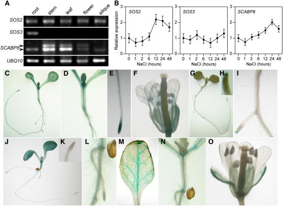
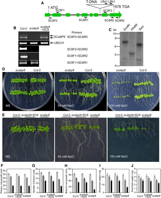
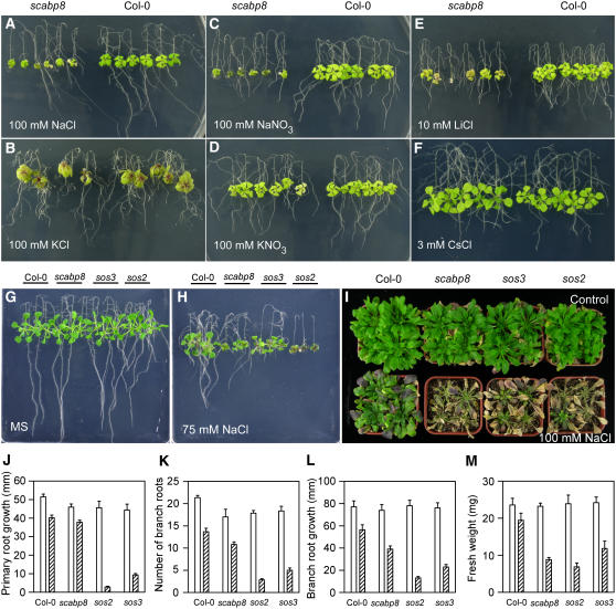
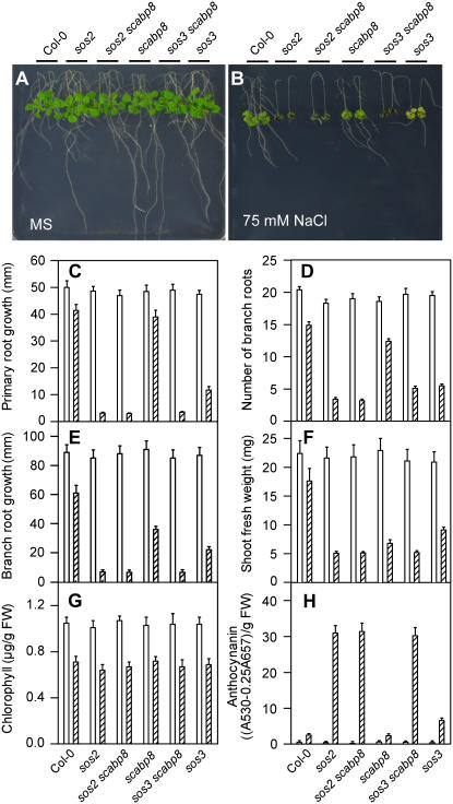
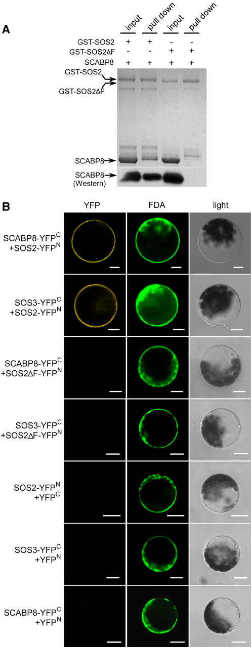
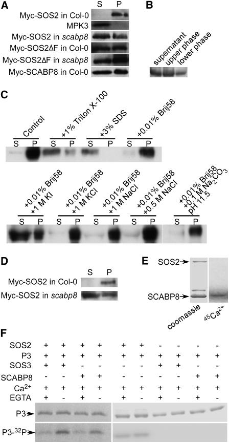
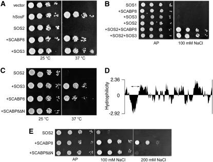
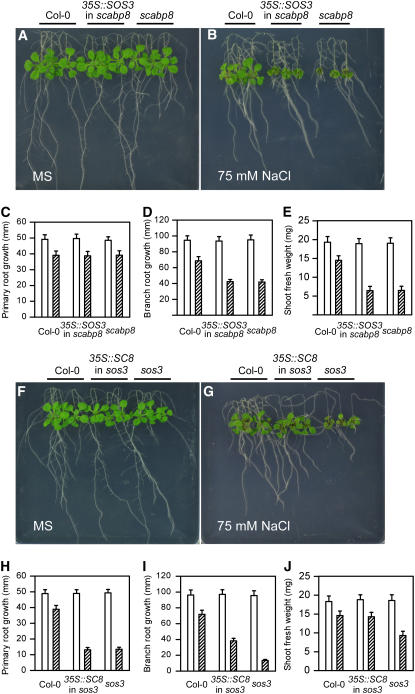
The SOS (for Salt Overly Sensitive) pathway plays essential roles in conferring salt tolerance in Arabidopsis thaliana. Under salt stress, the calcium sensor SOS3 activates the kinase SOS2 that positively regulates SOS1, a plasma membrane sodium/proton antiporter. We show that SOS3 acts primarily in roots under salt stress. By contrast, the SOS3 homolog SOS3-LIKE CALCIUM BINDING PROTEIN8 (SCABP8)/CALCINEURIN B-LIKE10 functions mainly in the shoot response to salt toxicity. While root growth is reduced in sos3 mutants in the presence of NaCl, the salt sensitivity of scabp8 is more prominent in shoot tissues. SCABP8 is further shown to bind calcium, interact with SOS2 both in vitro and in vivo, recruit SOS2 to the plasma membrane, enhance SOS2 activity in a calcium-dependent manner, and activate SOS1 in yeast. In addition, sos3 scabp8 and sos2 scabp8 display a phenotype similar to sos2, which is more sensitive to salt than either sos3 or scabp8 alone. Overexpression of SCABP8 in sos3 partially rescues the sos3 salt-sensitive phenotype. However, overexpression of SOS3 fails to complement scabp8. These results suggest that SCABP8 and SOS3 are only partially redundant in their function, and each plays additional and unique roles in the plant salt stress response.


Figures

Figure 1.
Figure 1.
Expression of SOS2, SOS3, and SCABP8 in Arabidopsis. (A) Expression of SOS2, SOS3, and SCABP8 in root, stem, leaf, flower, and silique determined by 30 cycles of RT-PCR with gene-specific primers. UBQ10 was a loading control. (B) Expression of SOS2, SOS3, and SCABP8 in Arabidopsis seedlings in response to salt stress. Error bars represent sd (n = 3). (C) to (F) SOS2 promoter-GUS expression in 10-d-old wild-type seedling (C), stem (D), root tip (E), and flower (F). (G) to (I) SOS3 promoter-GUS expression in 10-d-old wild-type seedling (G), root tip (H), and root (I). (J) to (O) SCABP8 promoter-GUS expression in 10-d-old wild-type seedling (J), root tip (K), root (L), leaf (M), stem (N), and flower (O).
Figure 2.
Figure 2.
scabp8 Is a Salt-Sensitive Mutant. (A) Structure of the SCABP8 gene. Filled green boxes indicate the exons, and the lines between boxes indicate introns. The primers used to identify T-DNA insertion and transcription of SCABP8 are marked with arrows. (B) Expression of SCABP8 in the wild type, the scabp8 mutant, and the complementary line. RT-PCR products were amplified from scabp8 with 30 cycles. The primer positions were identified in (A). UBQ10 was used as a loading control. (C) DNA gel blotting analysis of scabp8 genomic DNA probed with NPTII. (D) Five-day-old wild-type and scabp8 seedlings were transferred onto MS medium containing 0, 50, and 100 mM NaCl, respectively. The pictures were taken after 10 d of treatment. (E) Complementation of scabp8 by the wild-type SCABP8 gene. Five-day-old wild-type (left), transgenic scabp8 plants containing the wild-type SCABP8 gene (center), and scabp8 (right) seedlings were grown on MS medium with or without NaCl. (F) to (J) Primary root elongation (F), number of lateral roots (G), lateral root elongation (H), shoot fresh weight (I), and root fresh weight (J) were measured at day 10 after transfer. Open bars, without NaCl treatment; hatched bars, 50 mM NaCl treatment; closed bars, 100 mM NaCl treatment. Error bars represent sd (n > 15).
Figure 3.
Figure 3.
Na+ and Li+ Are the Toxic Ions to scabp8. (A) to (F) Five-day-old seedlings of the wild type and scabp8 were transferred onto MS medium with 100 mM NaCl (A), 100 mM KCl (B), 100 mM NaNO3 (C), 100 mM KNO3 (D), 10 mM LiCl (E), and 3 mM CsCl (F). The pictures were taken at 10 d after transfer. (G) to (I) Salt sensitivity of sos2, sos3, and scabp8 mutants. Five-day-old seedlings of the wild type, sos2, sos3, and scabp8 were transferred onto MS medium without (G) and with (H) 75 mM NaCl or transferred into soil (I). After 1 month of growth in soil, the plants were then treated with 100 mM NaCl for 2 weeks and photographed. (J) to (M) Primary root elongation (J), lateral root number (K), lateral root elongation (L), and fresh weight (M) regarding to (G) and (H) were measured at day 10 after transfer. Open bars, without NaCl treatment; hatched bars, 75 mM NaCl treatment. Error bars represent sd (n > 15).
Figure 4.
Figure 4.
Genetic Interaction of scabp8 with sos2 or sos3. Five-day-old seedlings of the wild type, sos2, sos2 scabp8, scabp8, scabp8 sos3, and sos3 were transferred onto MS medium without (A) and with (B) 75 mM NaCl. The pictures were taken at day 10. Primary root elongation (C), lateral root number (D), lateral root elongation (E), fresh shoot weight (FW) (F), chlorophyll (G), and anthocynanin contents (H) were measured at day 10 after transfer. Open bars, without NaCl treatment; hatched bars, 75 mM NaCl treatment. Error bars represent sd (n > 15).
Figure 5.
Figure 5.
Interaction of SOS2 with SOS3 and SCABP8. (A) SOS2 interacts with SCABP8. Lane 1, input of 1 μg of GST-SOS2 and 5 μg of SCABP8; lane 3, input of 1 μg of GST-SOS2ΔFISL and 5 μg of SCABP8; lanes 2 and lane 4, the pull-down products of lines 1 and 3 after washing four times. Protein gel blot analysis of 1% of the input proteins and pull-down proteins with anti-SCABP8 antibodies (bottom). (B) SOS2 interacts with SOS3 and SCABP8 in vivo. Different combinations of the N-YFP and C-YFP fusion constructs as indicated were cotransferred into Arabidopsis protoplasts. The images were collected under the Zeiss confocal microscope. Left, YFP signal; right, the light microscope pictures; middle, the protoplast stained with FDA (green). Bars = 10 μm.
Figure 6.
Figure 6.
Transient Expression of GFP-Tagged SCABP8 in Onion Epidermal Cells Visualized by Epifluorescence Microscopy. (A) Epifluorescence after 20 h of transient expression of SCABP8-GFP. (B) Epifluorescence after 40 h of transient expression of SCABP8-GFP. (C) Magnification of the cell shown in (B). PM, plasma membrane; cyt, cytosol; vac, vacuole. (D) Clear field image of cells plasmolized with 0.5 M sorbitol for 10 min. (E) Epifluorescence of the same cell shown in (D). (F) Fluorescence pattern of truncated SCABP8ΔN fused to GFP. (G) Fluorescence pattern of SCABP8ΔN-GFP. Bars = 50 μm.
Figure 7.
Figure 7.
SCABP8 Binds to Calcium, Increases SOS2 Activity, and Is Required for Association of SOS2 with the Plasma Membrane. (A) Myc-SOS2, -SOS2ΔF, and -SCABP8 plasmids were transferred into wild-type and scabp8 leaf protoplasts. The membrane fraction (P) and soluble fraction (S) were extracted from the protoplasts and probed with anti-myc or anti-MPK3 antibodies. (B) Vesicles from Arabidopsis overexpressing 35S:myc-SCABP8 were prepared by two-phase partitioning. Five micrograms of proteins from the soluble fraction, upper phase, and lower phase were subjected to protein gel blot analysis with anti-Myc antibody. (C) Plasma membrane–enriched vesicles were treated with 0.01% Brij58, 1% Triton X-100, 3% SDS, 1 M KI, 1 M KCl, 1 M NaCl, 0.5 M NaCl, and 0.1 M Na2CO3, pH 11.5, for 30 min. After centrifugation at 150,000g at 4°C for 60 min, equal aliquots of supernatants (S) and pellets (P) were loaded and detected with anti-Myc antibody. (D) Plasma membrane–enriched vesicles were purified by a two-phase method from protoplasts transformed with 35S:myc-SOS2, and 5 μg of soluble proteins (S) and 1 μg of plasma membrane–enriched proteins (P) were loaded and detected with anti-Myc. (E) SCABP8 binds calcium. GST-SOS2 and SCABP8 proteins were separated by SDS-PAGE and stained with Coomassie blue (left) or electroblotted onto a cellulose membrane, incubated with 45Ca2+, and autoradiographed (right). (F) Phosphorylation of GST-P3 by SOS2 kinase. Coomassie blue–stained GST-P3 in an SDS-PAGE gel (top); GST-P3 phosphorylation activity (bottom).
Figure 8.
Figure 8.
SCABP8 Recruits SOS2 to the Plasma Membrane and Activates SOS1 in Yeast. (A) Recruitment of SOS2 to the plasma membrane. A yeast cdc25-2 mutant was transformed with an empty pADNs plasmid (vector), a full-length human hSos protein as positive control (hSosF), and the hSos:SOS2 translational fusion (SOS2) in the same vector pADNs. Cells expressing hSos:SOS2 were cotransformed with plasmids directing the expression of SCABP8 (+SCABP8) or SOS3 (+SOS3). Five microliters of serial decimal dilutions were spotted onto YPD plates and incubated at 25 or 37°C for 3 d. (B) Activation of SOS1. Yeast AXT3K cells expressing SOS1 and transformed with the indicated combinations of Arabidopsis genes were grown overnight in liquid AP medium with 1 mM KCl. Five microliters of serial decimal dilutions were spotted onto plates of the same medium (AP) or supplemented with 100 mM NaCl. Plates were incubated at 28°C for 4 d. (C) Truncated SCABP8 does not recruit SOS2 to the plasma membrane. Mutant cdc25-2 expressing the hSos:SOS2 translational fusion (SOS2) was cotransformed with SOS3 (+SOS3), full-length SCABP8 (+SCABP8), or a truncated form of SCABP8 (+SCABP8ΔN) and processed as in (A). (D) Hydrophilicity plot of SCABP8. The internal deletion from residues Val-13 to Val-35 spanning the hydrophobic domain is indicated by the double arrow. (E) Truncated SCABP8 interacts with SOS2 to activate SOS1. Yeast AXT3K cells coexpressing SOS1 and SOS2 (SOS2) were transformed with wild-type SCABP8 (+SCABP8) or truncated SCABP8 (+SCABP8ΔN) and plated in AP medium with the indicated NaCl concentrations as in (B).
Figure 9.
Figure 9.
SCABP8 Partially Suppresses the sos3 Phenotype. (A) to (E) Five-old-day seedlings of the wild type (left), overexpression of SOS3 in scabp8 (center), and scabp8 (right) were transferred onto MS medium without (A) or with (B) 75 mM NaCl. Primary root elongation (C), lateral root elongation (D), and shoot fresh weight (E) were measured. (F) to (J) Five-old-day seedlings of the wild type (left), overexpression of SCABP8 in sos3 (center), and sos3 (right) were transferred onto MS medium without (F) or with (G) 75 mM NaCl. Primary root elongation (H), lateral root elongation (I), and shoot fresh weight (J) were measured. The pictures were taken at day 15. Open bars, without NaCl treatment; hatched bars, 75 mM NaCl treatment. Error bars represent sd (n > 15).

Similar articles

Cited by

References

    1. Albrecht, V., Weinl, S., Blazevic, D., D'Angelo, C., Batistic, O., Kolukisaoglu, U., Bock, R., Harter, K., and Kudla, J. (2003). The calcium sensor CBL1 integrates plant responses to abiotic stresses. Plant J. 36 457–470. - PubMed
    1. Alonso, J.M., et al. (2003). Genome-wide insertional mutagenesis of Arabidopsis thaliana. Science 301 653–657. - PubMed
    1. Aronheim, A., Zandi, E., Hennemann, H., Elledge, S.J., and Karin, M. (1997). Isolation of an AP-1 repressor by a novel method for detecting protein-protein interactions. Mol. Cell. Biol. 17 3094–3102. - PMC - PubMed
    1. Batistic, O., and Kudla, J. (2004). Integration and channeling of calcium signaling through the CBL calcium sensor/CIPK protein kinase network. Planta 219 915–924. - PubMed
    1. Cheng, N.-H., Pittman, J.K., Zhu, J.-K., and Hirschi, K.D. (2004). The protein kinase SOS2 activates the Arabidopsis H+/Ca2+ antiporter CAX1 to integrate calcium transport and salt tolerance. J. Biol. Chem. 279 2922–2926. - PubMed

Publication types

MeSH terms