. 2007 Apr;22(2):183-91.
doi: 10.3346/jkms.2007.22.2.183.

Time sequence of airway remodeling in a mouse model of chronic asthma: the relation with airway hyperresponsiveness

Affiliations

Time sequence of airway remodeling in a mouse model of chronic asthma: the relation with airway hyperresponsiveness

Seung Joon Kim et al. J Korean Med Sci. 2007 Apr.

Abstract

During the course of establishing an animal model of chronic asthma, we tried to elucidate the time sequence of airway hyperresponsiveness (AHR), airway inflammation, airway remodeling, and associated cytokines. Seven-week-old female BALB/c mice were studied as a chronic asthma model using ovalbumin (OVA). After sensitization, mice were exposed twice weekly to aerosolized OVA, and were divided into three groups depending on the duration of 4 weeks, 8 weeks, and 12 weeks. At each time point, airway responsiveness, inflammatory cells, cytokines in bronchoalveolar lavage fluids (BALF), serum OVA-specific IgE, IgG1, IgG2a, and histological examination were carried out. AHR to methacholine, increased levels of OVA-specific IgG1 and IgG2a, and goblet cell hyperplasia were continuously sustained at each time point of weeks. In contrast, we observed a time-dependent decrease in serum OVA-specific IgE, BALF eosinophils, BALF cytokines such as IL-13, transforming growth factor-beta1, and a time-dependent increase in BALF promatrix metalloproteinase-9 and peribronchial fibrosis. In this OVA-induced chronic asthma model, we observed airway remodelings as well as various cytokines and inflammatory cells being involved in different time-dependent manners. However, increased airway fibrosis did not directly correlate with a further increase in airway hyperresponsiveness.


Figures

Fig. 1
Fig. 1
Schematic representation of systemic sensitization and provocation protocol in each group of mouse asthma model. IP, intraperitoneal injection.
Fig. 2
Fig. 2
Airway hyperresponsiveness to methacholine in each group of mouse asthma model. Compared with the control group, asthma groups showed significantly increased airway hyperresponsiveness for 12 weeks (*p<0.01 vs. control group).
Fig. 3
Fig. 3
Changes of inflammatory cells in bronchoalveolar lavage fluid in each group of mouse asthma model. Compared with the control group, asthma groups showed significantly increased inflammatory cells including eosinophils, lymphocytes and neutrophils (*p<0.01). In the asthma groups, eosinophils were significantly decreased after prolonged OVA inhalations for 8 weeks and 12 weeks p<0.01).
Fig. 4
Fig. 4
Photomicrographs of MMP-9 (I) and TIMP-1 (II) immunoreactivity in the bronchoalveolar lavage cells of each group of mouse asthma model (×400): (A) Control group, (B) Group I (4 weeks OVA inhalation), (C) Group II (8 weeks OVA inhalation), (D) Group III (12 weeks OVA inhalation). Positive staining cells expressed blue color in their cytoplasm. Compared with the control group, MMP-9 and TIMP-1 expressions were obvious in asthma groups.
Fig. 5
Fig. 5
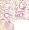
Photomicrographs of PAS stain of lung tissue in each group of mouse asthma model (×100): (A) Control group, (B) Group I (4 weeks OVA inhalation), (C) Group II (8 weeks OVA inhalation), (D) Group III (12 weeks OVA inhalation). Compared with the control group, asthma groups showed increased goblet cell hyperplasia, and furthermore airway obstructions were developed due to mucus secretion and fibrosis of the bronchial wall (E).
Fig. 6
Fig. 6
Changes of goblet cell hyperplasia in each group of mouse asthma model. The hyperplasia of goblet cells in the epithelial lining was expressed by a score according to the percentage of the goblet cells in the epithelial cells: grade 0, no goblet cells; grade 1, <25%; grade 2, 25-50%; grade 3, 51-75%; grade 4, >75% or total bronchial obstruction due to mucus. Compared with the control group, asthma groups showed significantly increased goblet cell hyperplasia (*p<0.01). However, there were no significant differences between asthma groups.
Fig. 7
Fig. 7
Photomicrographs of Masson's trichrome stain of lung tissue in each group of mouse asthma model (×100): (A) Control group, (B) Group I (4 weeks OVA inhalation), (C) Group II (8 weeks OVA inhalation), (D) Group III (12 weeks OVA inhalation). Peribronchial fibrosis developed after prolonged OVA inhalations.
Fig. 8
Fig. 8
Changes of peribronchial collagen deposition in each group of mouse asthma model. Compared with the control group, asthma groups showed significantly increased peribronchial fibrosis (*p<0.01). In the asthma groups, Group II showed more significant increase of peribronchial fibrosis than Group I (p<0.01), and Group III showed more significant increase of peribronchial fibrosis than Group II (p<0.05).

References

    1. Brown PJ, Greville HW, Finucane KE. Asthma and irreversible airflow obstruction. Thorax. 1984;39:131–136. - PMC - PubMed
    1. Broide DH, Lotz M, Cuomo AJ, Coburn DA, Federman EC, Wasserman SI. Cytokines in symptomatic asthma airways. J Allergy Clin Immunol. 1992;89:958–967. - PubMed
    1. Jeffery PK, Wardlaw AJ, Nelson FC, Collins JV, Kay AB. Bronchial biopsies in asthma. An ultrastructural, quantitative study and correlation with hyperreactivity. Am Rev Respir Dis. 1989;140:1745–1753. - PubMed
    1. Bousquet J, Chanez P, Lacoste JY, White R, Vic P, Godard P, Michel FB. Asthma: a disease remodeling the airways. Allergy. 1992;47:3–11. - PubMed
    1. Redington AE, Howarth PH. Airway wall remodelling in asthma. Thorax. 1997;52:310–312. - PMC - PubMed