Comparative Study
. 2007 May;39(5):614-22.
doi: 10.1038/ng2031. Epub 2007 Apr 22.

Modifiers of epigenetic reprogramming show paternal effects in the mouse

Affiliations
Comparative Study

Modifiers of epigenetic reprogramming show paternal effects in the mouse

Suyinn Chong et al. Nat Genet. 2007 May.

Abstract

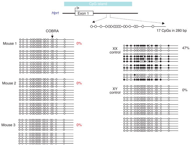
There is increasing evidence that epigenetic information can be inherited across generations in mammals, despite extensive reprogramming both in the gametes and in the early developing embryo. One corollary to this is that disrupting the establishment of epigenetic state in the gametes of a parent, as a result of heterozygosity for mutations in genes involved in reprogramming, could affect the phenotype of offspring that do not inherit the mutant allele. Here we show that such effects do occur following paternal inheritance in the mouse. We detected changes to transcription and chromosome ploidy in adult animals. Paternal effects of this type have not been reported previously in mammals and suggest that the untransmitted genotype of male parents can influence the phenotype of their offspring.


Conflict of interest statement

COMPETING INTERESTS STATEMENT

The authors declare no competing financial interests.

Figures

Figure 1
Figure 1
Momme D4 is caused by a mutation in Smarca5, which encodes the ISWI chromatin remodeler Snf2h. (a) A single–base pair mutation (a T-to-A transversion) occurs in exon 12 of Smarca5 in homozygous Momme D4 embryos (MD4/MD4). (b) Schematic of the Snf2h protein structure. The point mutation in Momme D4 causes a nonconservative amino acid substitution of a large aromatic residue (tryptophan) with a basic residue (arginine). This residue is highly conserved in ISWI members across species. (c) RNA blot analysis performed on poly(A)+ mRNA does not show any difference in Smarca5 mRNA levels or transcript size between homozygous (MD4/MD4), heterozygous (MD4/+) and wild-type (+/+) individuals. Membranes were stripped and hybridized with mouse Gapdh. (d) Whole-cell lysate from 17.5-d.p.c. homozygous (MD4/MD4), heterozygous (MD4/+) and wild-type (+/+) Momme D4 embryos, probed with anti-Snf2h (top row). Membranes were also probed with anti-γ-tubulin (bottom row). (e) FACS data of offspring from a cross between Momme D4 heterozygotes and a Smarca5+/− knockout allele. The GFP transgene expression profile of erythrocytes from 3-week-old mice is shown. The x axis represents the erythrocyte fluorescence on a logarithmic scale, and the y axis is the number of cells detected at each fluorescence level.
Figure 2
Figure 2
Coat-color phenotypes after paternal transmission of Momme D4. (a) Isogenic mice carrying the agouti viable yellow allele show different coat colors (yellow, mottled and agouti) depending on the epigenetic state of the LTR promoter. (b) Pedigree produced from a Smarca5MommeD4/+ sire and yellow Avy/a dam. Offspring heterozygous for the Momme D4 mutation demonstrated a shift in penetrance at Avy toward mottled when compared with wild-type littermates (P < 0.005) (a subset of this data is shown in ref. 5). (c) The coat color phenotypes of wild-type offspring from a wild-type sire were compared with the wild-type offspring from a Smarca5MommeD4/+ sire. Both sets of offspring were genetically identical (Smarca5+/+; Avy/A); the only difference was that one group originated from gametes produced in a Smarca5MommeD4/+ sire. Wild-type offspring from Smarca5MommeD4/+ sires showed a significant increase in the proportion of animals with yellow coats when compared with the wild-type offspring from wild-type sires.
Figure 3
Figure 3
Localization of Snf2h during spermatogenesis. (a) Preabsorbed control to indicate the specificity of immunohistochemical staining. (b,c) Snf2h protein was present in prophase I spermatocytes to stage VII pachytene spermatocytes and in terminally differentiating elongating spermatids. All other germ cell types and Sertoli cells were unstained. However, we observed Snf2h staining in the cytoplasm of Leydig cells within the interstitial space. Z = zygotene spermatocyte, P = pachytene spermatocyte, r = round spermatid, E = elongated spermatid. Scale bars = 100 μm.
Figure 4
Figure 4
Momme D2 is caused by a mutation in Dnmt1. (a) A single–base pair mutation (C-to-A transversion) occurs in exon 25 of Dnmt1 in heterozygous Momme D2 mice. (b) A schematic of the Dnmt1 protein structure. The point mutation in Momme D2 causes a nonconservative amino acid substitution in which a small polar residue (threonine) is replaced with a larger, basic residue (lysine). This residue is highly conserved in Dnmt1 orthologs. (c) Homology mapping, generated using Swiss-Model and the coordinates of the proximal bromo-adjacent homology (BAH) domain of the BAH1 protein (PDB code 1W4S) shows the relevant amino acid in the center of the BAH domain. (d) Nuclear lysate from individual 9.5-d.p.c. wild-type embryos (+/+), heterozygous embryos (MD2/+) and four pooled homozygous (MD2/MD2) embryos, probed with anti-Dnmt1 (top row). As a loading control, blots were probed with anti-γ-tubulin (bottom row). (e) Coat color phenotypes following paternal transmission of Momme D2. Pedigree produced from a Dnmt1MommeD2/+ sire and a yellow Avy dam. Wild-type offspring from a Dnmt1MommeD2/+ sire were more likely to be yellow than wild-type offspring from a wild-type sire. Both sets of offspring were genetically identical (Dnmt1+/+; Avy/A), except that one group originated from gametes produced in a Dnmt1MommeD2/+ sire. (f) Coat-color phenotypes following paternal transmission of Momme D3. Pedigree produced from a Momme D3+/− sire and a yellow Avy dam showed no paternal effects.
Figure 5
Figure 5
Hypomethylation of the X-linked Hprt CpG island in adult females from Dnmt3l+/− sires. Bisulfite sequence analysis of a 280-bp region located within intron 1 of the mouse Hprt locus, encompassing 17 CpG dinucleotides. Methylated (filled circles) and unmethylated (open circles) CpG dinucleotides are shown for a number of independently sequenced templates (horizontal lines). Each group of horizontal lines represents an independent bisulfite conversion and PCR reaction. The percentage of methylation in each mouse is shown (calculated from the number of methylated CpGs divided by the total CpGs sequenced, multiplied by 100). Clones were included in the samples only if they could be distinguished from others in the sample by non-CpG methylation. Any clones with >5% non-CpG methylation (an indication of incomplete bisulfite conversion) were excluded from the data set.
Figure 6
Figure 6
A single sex chromosome in an adult female from a Dnmt3l+/− sire. Array comparative genomic hybridization (CGH) analysis of mouse 2. The genome view (left) and expanded X chromosome view (right) were created using Agilent CGH analytics software. The genome view shows an ideogram of each mouse chromosome, with a dotted vertical line beside each ideogram. Horizontal bars that extend to the right and left of this dotted line indicate gain and loss calls, respectively, by the software. The chromosome view shows a moving average plot with a 0.5-Mb window. The moving average is shifted from 0 to −1 along the entire X chromosome, indicating a single X chromosome.
Figure 7
Figure 7
A single sex chromosome in an F1 hybrid embryo from a Dnmt3l+/− sire. Array CGH analysis of a mouse with paternal X chromosome only (see Table 1). The genome view (left) and expanded X chromosome view (right) were created using Agilent CGH analytics software. The genome view shows an ideogram of each mouse chromosome, with a dotted vertical line beside each ideogram. Horizontal bars that extend to the right and left of this dotted line indicate gain and loss calls, respectively, by the software. The chromosome view shows a moving average plot with a 0.5-Mb window. In this mouse, the moving average is shifted from 0 to −1 along the entire X chromosome, indicating a single X chromosome.

Similar articles

Cited by

References

    1. Chong S, Whitelaw E. Epigenetic germline inheritance. Curr Opin Genet Dev. 2004;14:692–696. - PubMed
    1. Rakyan V, Whitelaw E. Transgenerational epigenetic inheritance. Curr Biol. 2003;13:R6. - PubMed
    1. Fitch KR, Yasuda GK, Owens KN, Wakimoto BT. Paternal effects in Drosophila: implications for mechanisms of early development. Curr Top Dev Biol. 1998;38:1–34. - PubMed
    1. Payer B, et al. Stella is a maternal effect gene required for normal early development in mice. Curr Biol. 2003;13:2110–2117. - PubMed
    1. Blewitt ME, et al. An N-ethyl-N-nitrosourea screen for genes involved in variegation in the mouse. Proc Natl Acad Sci USA. 2005;102:7629–7634. - PMC - PubMed

Publication types

MeSH terms

Associated data