

Gallstone ileus: report of two cases and review of the literature
- PMID: 17451220
- PMCID: PMC4147014
- DOI: 10.3748/wjg.v13.i8.1295
Gallstone ileus: report of two cases and review of the literature
Abstract
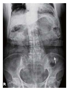
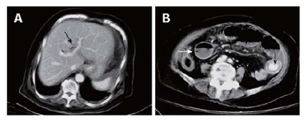
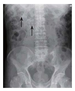
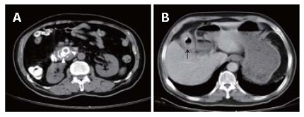
Gallstone ileus is a rare disease and accounts for 1%-4% of all cases of mechanical intestinal obstruction. It usually occurs in the elderly with a female predominance and may result in a high mortality rate. Its diagnosis is difficult and early diagnosis could reduce the mortality. Surgery remains the mainstay of treatment. We report two cases of gallstone ileus. The first was a 78-year old woman who had a 2-d history of vomiting and epigastralgia. Plain abdominal film suggested small bowel obstruction clinically attributed to adhesions. Later on, gallstone ileus was diagnosed by abdominal computed tomography (CT) based on the presence of pneumobilia, bowel obstruction, and an ectopic stone within the jejunum. She underwent emergent laparotomy with a one-stage procedure of enterolithotomy, cholecystectomy and fistula repair. The second case was a 76-year old man with a 1-wk history of epigastralgia. Plain abdominal film showed two round calcified stones in the right upper quadrant. Fistulography confirmed the presence of a cholecystoduodenal fistula and gallstone ileus was also diagnosed by abdominal CT. We attempted to remove the stones endoscopically, but failed leading to an emergent laparotomy and the same one-stage procedure as for the first case. The postoperative courses of the two cases were uneventful. Inspired by these 2 cases we reviewed the literature on the cause, diagnosis and treatment of gallstone ileus.
Figures
References
-
- Richards WO, Williams LF. Obstruction of the large and small intestine. Surg Clin North Am. 1988;68:355–376. - PubMed
-
- Abou-Saif A, Al-Kawas FH. Complications of gallstone disease: Mirizzi syndrome, cholecystocholedochal fistula, and gallstone ileus. Am J Gastroenterol. 2002;97:249–254. - PubMed
-
- Kurtz RJ, Heimann TM, Kurtz AB. Gallstone ileus: a diagnostic problem. Am J Surg. 1983;146:314–317. - PubMed
-
- Clavien PA, Richon J, Burgan S, Rohner A. Gallstone ileus. Br J Surg. 1990;77:737–742. - PubMed
-
- Reisner RM, Cohen JR. Gallstone ileus: a review of 1001 reported cases. Am Surg. 1994;60:441–446. - PubMed
Publication types
MeSH terms
LinkOut - more resources
Full Text Sources
