

Characterization of neuronal subsets surrounded by perineuronal nets in the rhesus auditory brainstem
- PMID: 17451528
- PMCID: PMC2375744
- DOI: 10.1111/j.1469-7580.2007.00713.x
Characterization of neuronal subsets surrounded by perineuronal nets in the rhesus auditory brainstem
Abstract
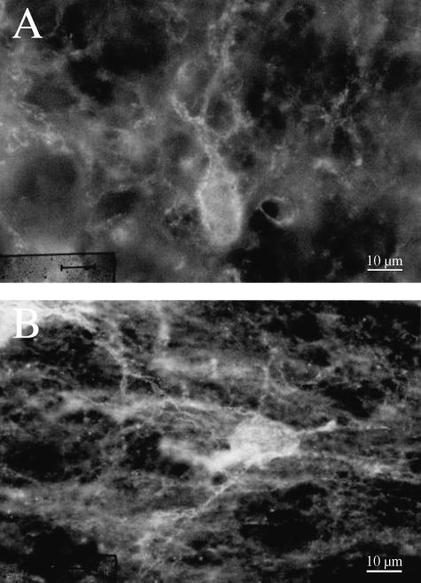
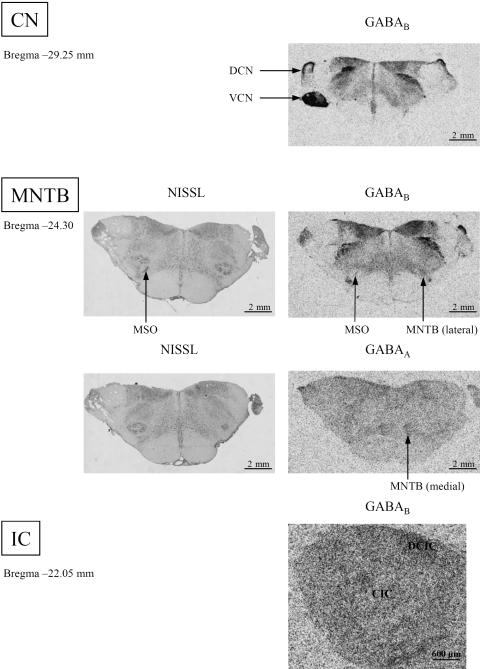
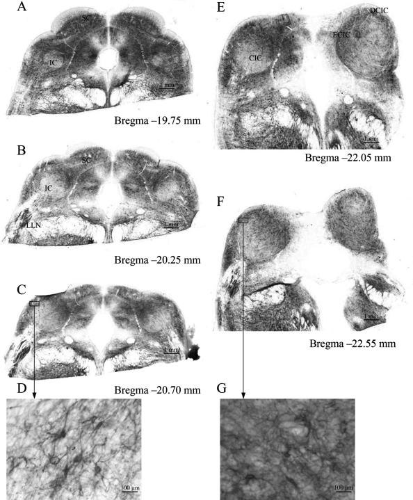
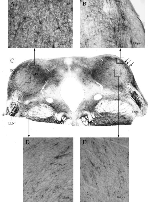
The distribution of perineuronal nets and the potassium channel subunit Kv3.1b was studied in the subdivisions of the cochlear nucleus, the medial nucleus of the trapezoid body, the medial and lateral superior olivary nuclei, the lateral lemniscal nucleus and the inferior colliculus of the rhesus monkey. Additional sections were used for receptor autoradiography to visualize the patterns of GABAA and GABAB receptor distribution. The Kv3.1b protein and perineuronal nets [visualized as Wisteria floribunda agglutinin (WFA) binding] were revealed, showing corresponding region-specific patterns of distribution. There was a gradient of labelled perineuronal nets which corresponded to that seen for the intensity of Kv3.1b expression. In the cochlear nucleus intensely and faintly stained perineuronal nets were intermingled, whereas in the medial nucleus of the trapezoid body the pattern changed to intensely stained perineuronal nets in the medial part and weakly labelled nets in its lateral part. In the inferior colliculus, intensely labelled perineuronal nets were arranged in clusters and faintly labelled nets were arranged in sheets. Using receptor autoradiography, GABAB receptor expression in the anterior ventral cochlear nucleus was revealed. The medial part of the medial nucleus of the trapezoid body showed a high number of GABAA binding sites whereas the lateral part exhibited more binding sites for GABAB. In the inferior colliculus, we found moderate GABAB receptor expression. In conclusion, intensely WFA-labelled structures are those known to be functionally involved in high-frequency processing.
Figures

Similar articles
-
Distribution of perineuronal nets in the human superior olivary complex.Hear Res. 2010 Jun 14;265(1-2):15-24. doi: 10.1016/j.heares.2010.03.077. Epub 2010 Mar 20. Hear Res. 2010. PMID: 20307636
-
In vivo and in vitro labelling of perineuronal nets in rat brain.Brain Res. 1996 May 13;720(1-2):84-92. doi: 10.1016/0006-8993(96)00152-7. Brain Res. 1996. PMID: 8782900
-
Cortical neurons immunoreactive for the potassium channel Kv3.1b subunit are predominantly surrounded by perineuronal nets presumed as a buffering system for cations.Brain Res. 1999 Sep 18;842(1):15-29. doi: 10.1016/s0006-8993(99)01784-9. Brain Res. 1999. PMID: 10526091
-
Localization of two high-threshold potassium channel subunits in the rat central auditory system.J Comp Neurol. 2001 Aug 20;437(2):196-218. doi: 10.1002/cne.1279. J Comp Neurol. 2001. PMID: 11494252
-
Perineuronal nets in the auditory system.Hear Res. 2015 Nov;329:21-32. doi: 10.1016/j.heares.2014.12.012. Epub 2015 Jan 9. Hear Res. 2015. PMID: 25580005 Review.
Cited by
-
The mammalian interaural time difference detection circuit is differentially controlled by GABAB receptors during development.J Neurosci. 2010 Jul 21;30(29):9715-27. doi: 10.1523/JNEUROSCI.1552-10.2010. J Neurosci. 2010. PMID: 20660254 Free PMC article.
-
VGLUT1 and VGLUT2 mRNA expression in the primate auditory pathway.Hear Res. 2011 Apr;274(1-2):129-41. doi: 10.1016/j.heares.2010.11.001. Epub 2010 Nov 24. Hear Res. 2011. PMID: 21111036 Free PMC article. Review.
-
Deafferentation-induced redistribution of MMP-2, but not of MMP-9, depends on the emergence of GAP-43 positive axons in the adult rat cochlear nucleus.Neural Plast. 2011;2011:859359. doi: 10.1155/2011/859359. Epub 2011 Oct 23. Neural Plast. 2011. PMID: 22135757 Free PMC article.
-
Perineuronal nets and GABAergic cells in the inferior colliculus of guinea pigs.Front Neuroanat. 2014 Jan 8;7:53. doi: 10.3389/fnana.2013.00053. eCollection 2014. Front Neuroanat. 2014. PMID: 24409124 Free PMC article.
-
Wisteria Floribunda Agglutinin-Labeled Perineuronal Nets in the Mouse Inferior Colliculus, Thalamic Reticular Nucleus and Auditory Cortex.Brain Sci. 2016 Apr 13;6(2):13. doi: 10.3390/brainsci6020013. Brain Sci. 2016. PMID: 27089371 Free PMC article.
References
-
- Aitkin LM, Webster WR, Veale JL, Crosby DC. Inferior colliculus. I. Comparison of response properties of neurons in central, pericentral, and external nuclei of adult cat. J Neurophysiol. 1975;38:1196–1207. - PubMed
-
- Aitkin L, Schuck D. Low frequency neurons in the lateral central nucleus of the cat inferior colliculus receive their input predominantly from the medial superior olive. Hear Res. 1985;17:87–93. - PubMed
-
- Aitkin L, Tran L, Syka J. The responses of neurons in subdivisions of the inferior colliculus of cats to tonal, noise and vocal stimuli. Exp Brain Res. 1994;98:53–64. - PubMed
-
- Atoji Y, Yamamoto Y, Suzuki Y, Matsui F, Oohira A. Immunohistochemical localization of neurocan in the lower auditory nuclei of the dog. Hear Res. 1997;110:200–208. - PubMed
-
- Bazwinsky I, Hilbig H, Bidmon HJ, Rübsamen R. Characterization of the human superior olivary complex by calcium binding proteins and neurofilament H (SMI-32) J Comp Neurol. 2003;456:292–303. - PubMed
MeSH terms
Substances
LinkOut - more resources
Full Text Sources
