. 2007 May;210(5):542-54.
doi: 10.1111/j.1469-7580.2007.00723.x.

Skeletogenesis in the swell shark Cephaloscyllium ventriosum

Affiliations

Skeletogenesis in the swell shark Cephaloscyllium ventriosum

B Frank Eames et al. J Anat. 2007 May.

Abstract

Extant chondrichthyans possess a predominantly cartilaginous skeleton, even though primitive chondrichthyans produced bone. To gain insights into this peculiar skeletal evolution, and in particular to evaluate the extent to which chondrichthyan skeletogenesis retains features of an osteogenic programme, we performed a histological, histochemical and immunohistochemical analysis of the entire embryonic skeleton during development of the swell shark Cephaloscyllium ventriosum. Specifically, we compared staining properties among various mineralizing tissues, including neural arches of the vertebrae, dermal tissues supporting oral denticles and Meckel's cartilage of the lower jaw. Patterns of mineralization were predicted by spatially restricted alkaline phosphatase activity earlier in development. Regarding evidence for an osteogenic programme in extant sharks, a mineralized tissue in the perichondrium of C. ventriosum neural arches, and to a lesser extent a tissue supporting the oral denticle, displayed numerous properties of bone. Although we uncovered many differences between tissues in Meckel's cartilage and neural arches of C. ventriosum, both elements impart distinct tissue characteristics to the perichondral region. Considering the evolution of osteogenic processes, shark skeletogenesis may illuminate the transition from perichondrium to periosteum, which is a major bone-forming tissue during the process of endochondral ossification.


Figures
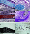
Fig. 1
Fig. 1
Typical histological and immunohistochemical properties of vertebrate bone and cartilage. See Materials and methods for specific histological dye affinities. (A) HBQ staining of cartilage (c) and perichondral bone (b) in ceratobranchial of E15 chick embryo. (B) HBQ staining of cartilage and dermal bone in Meckel's cartilage and dentary of E14 mouse embryo. (C) Safranin O staining of ceratobranchial in E12 chick embryo. (D) Trichrome staining of Meckel's cartilage and suprangular in E11 duck embryo. (E) Col2 immunostaining of quadrate in E9 chick embryo. (F) Col1 immunostaining of suprangular in E9 chick embryo. Abbreviations: b = bone; c = cartilage. Scale bars: A,C,D = 200 µm; B,E,F = 100 µm.
Fig. 2
Fig. 2
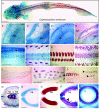
Exoskeletal and endoskeletal mineralization during swell shark ontogeny. (A) Dorsal view of 15-cm swell shark, stained with Alcian blue and Alizarin red. (B–E) Ventral views of swell shark lower jaws, anterior to top. Meckel's cartilage (Mk) stained with Alcian blue in 5-cm (B), 9-cm (C), 12-cm (D) and 15-cm (E) embryos. Alizarin red stained teeth (t) in 9-cm, 12-cm and 15-cm oral surfaces (C–E), and dermal denticles (dd) on 15-cm head (E). (F–I) Dorsal views of swell shark spines, anterior to left. Vertebrae (v) stained with Alcian blue in 5-cm (F), 9-cm (G) and 15-cm (I) embryos; note slight Alcian blue staining of dermal elements in 5-cm embryo (F, arrowhead). Vertebrae stained for mineralization in 9-cm embryos (H) that did not undergo acid alcohol treatment (compare with G), and in 15-cm embryos (I). Alizarin red staining also marked development of dermal denticles along the dorsal surface of the spine in 9-cm (G) and 15-cm embryos (I); dermal denticles were removed from the sample in H. (J–L) Lateral views of swell shark caudal fins, dorsal to top, anterior to left. First signs of mineralization detected in 5-cm embryos (J,K); the caudal dermal denticles demonstrate a different morphology than either the initial rows along the dorsal surface or those that eventually cover the body (compare K to E, I and L), and they persist in 15-cm embryos (L). (M–Q) Transverse views of swell shark vertebrae, dorsal to left. The 15-cm embryo (M) illustrates three portions of the vertebrum: neural arches (na), centrum (c) and hemal arches (ha). Alcian blue stained neural arches of 9-cm embryo (N). Alizarin red staining was observed in neural arches of a 9-cm embryo that did not undergo acid alcohol treatment (O). Txined with Alcian blue and an outer tissue (black arrowhead) that stained with Alizarin red. Three distinct ringed layers of the centrum (c) in a 15-cm embryo (Q) were visible: inner and outer layers stained with Alcian blue, while a middle layer stained for mineralization. Abbreviations: c = centrum; dd = dermal denticle; ha = hemal arch; Mk = Meckel's cartilage; na = neural arch; v = vertebrum. Scale bars: A = 1 cm; B–D, F–H = 1 mm; E, I, J, L, M = 2 mm; K, N–Q = 0.5 mm.
Fig. 5
Fig. 5
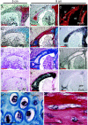
Shark Meckel's cartilage development and mineralization. (A–H) Sagittal sections of lower jaws in 5-cm (A,C,E,G) and 9-cm (B,D,F,H) swell shark embryos. Meckel's cartilage (Mk) stained with Safranin O and Alcian blue in 5-cm (A,C) and 9-cm (B,D) embryos. No staining of Meckel's cartilage with direct red of the HBQ protocol was observed in 5-cm (C) or 9-cm (D) embryos. Chondrocyte hypertrophy was apparent in Meckel's cartilage of the 9-cm embryo (B, arrows). No Aniline blue staining of the Trichrome protocol was observed in Meckel's cartilage of 5-cm (E) or 9-cm (F) embryos. No alkaline phosphatase activity was discerned in Meckel's cartilage of the 5-cm embryo (G). Intense alkaline phosphatase activity was evident in discrete groups of cells (arrowheads) near the surface of Meckel's cartilage in the 9-cm embryo (H); note alkaline phosphatase activity in the developing tooth (H, arrow). (I) Ventral view of lower jaw in 15-cm swell shark embryo. Superficial Alizarin red staining was apparent in Meckel's cartilage of a 15-cm embryo (I). Abbreviations: AP = alkaline phosphatase; Mk = Meckel's cartilage; SafO = Safranin O; Tri = Trichrome. Scale bars: A–H = 200 µm; I = 1 mm.
Fig. 3
Fig. 3
A bone-like tissue in neural arches of the shark vertebrae. (A–N) Transverse sections of swell shark neural arches in 5-cm (A,D,G,J), 9-cm (B,C,E,F,H,I,K,L) and 15-cm (M,N) embryos. Cells producing a matrix that stained with Safranin O (A) and Alcian blue (D) projected dorsally from the centrum (c) around the neural tube (nt), forming the neural arch (na), in 5-cm embryos. The neural arch of 9-cm embryos had extended dorsally to encompass completely the neural tube, and contained two tissue layers; in addition to a central layer (cl) that stained with Safranin O, a surrounding layer (sl) did not stain with Safranin O (B). A tangential section of the 9-cm neural arch demonstrated that the central layer (of which only the most dorsal and ventral aspects were represented in this section) stained with Alcian blue, while the surrounding layer stained with direct red (E). Neither Aniline blue staining in the Trichrome protocol (G) nor alkaline phosphatase activity (J) was observed in the neural arch of 5-cm embryos. Aniline blue staining was strong only in the surrounding layer of the neural arch in the 9-cm embryo (H). Evidence of alkaline phosphatase was strong in both the central and the surrounding layers of the neural arch in the 9-cm embryo (K). Picro Sirius-stained neural arches of 9-cm embryos indicated that the surrounding layer contained tight bundling of collagen fibres, as revealed by red birefringence under polarized light; by contrast, the central layer showed loose collagen organization (C). The central layer of the 9-cm neural arch demonstrated strong immunoreactivity for Collagen type 2 (Col2), while the surrounding layer was Col2-negative (F). The central layer of the 9-cm neural arch showed no immunoreactivity for Collagen type 1 (Col1), while the surrounding layer was Col1-positive (I). An antibody for the precursor peptide of Col1 (pre-Col1) was reactive only at the dorsal aspect of the surrounding layer in the neural arch of the 9-cm embryo (L). High-magnifications of swell shark neural arches in 15-cm embryos revealed rounded, well-separated cells in the central layer (M) and flattened, more closely spaced cells in the surrounding layer (N). Abbreviations: AP = alkaline phosphatase; c = centrum; cl = central layer; Col1 = Collagen type 1; Col2 = Collagen type 2; na = neural arch; nt = neural tube; pre-Col1 = precursor peptide of Col1; SafO = Safranin O; sl = surrounding layer; Tri = Trichrome. Scale bars: A,D,G,J = 200 µm; B,C,E,F,H,I,K,L = 400 µm; M,N = 20 µm.
Fig. 4
Fig. 4
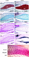
A bone-like tissue supporting the shark tooth. (A–H) Sagittal sections through the distal tip of the lower jaw in 15-cm swell shark embryo. Low (A) and higher magnification (B–F; black box in A) views revealed a tissue (arrow) supporting the tooth (t) that stained with direct red of the HBQ protocol (A,B) and Aniline blue of the Trichrome protocol (C). This tissue did not stain with Alcian blue (A,B) or Safranin O (D), but demonstrated alkaline phosphatase activity (E). While the tooth-supporting tissue showed some immunoreactivity to Collagen type 1 (F), high-magnification views (G,H; white box in F) of this tissue (outlined) in Picro Sirius-stained sections revealed that the collagen fibrils were not tightly wound. H is view of tissue in G under polarized light. Abbreviations: AP = alkaline phosphatase; Col1 = Collagen type 1 antibody; Mk = Meckel's cartilage; SafO = Safranin O; t = tooth; Tri = Trichrome. Scale bars: A = 800 µm; B–F = 200 µm; G,H = 100 µm.
Fig. 6
Fig. 6
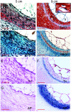
A bone-like tissue in the shark vertebral centrum. (A–H) Transverse sections of swell shark centra in 5-cm (A,C,E,G) and 9-cm (B,D,F,H) embryos. Safranin O staining (A) and Alcian blue of the HBQ protocol (C) distinguished two tissue layers in the centrum of vertebrae in the 5-cm shark embryo: lighter, more diffuse staining of an inner layer (il) of tissue adjacent to the notochord (n), and deeper, more concentrated staining in an outer layer (ol) of tissue. In 9-cm embryos, the centrum was divided into three tissue layers: inner (il) and outer (ol) layers stained with Safranin O, while a middle layer (ml) remained unstained by Safranin O (B). Note hypertrophic cells in the inner layer of the centrum (inset B′, which is a high-magnification of cells in the boxed area in B). Similar to Safranin O staining of the 9-cm centrum, Alcian blue stained inner and outer layers, but the middle layer stained slightly with direct red of the HBQ protocol (D). No Aniline blue staining in the Trichrome protocol was apparent in the 5-cm centrum (E), while only the middle layer of the 9-cm centrum stained slightly with Aniline blue (F). No alkaline phosphatase activity was present in the 5-cm centrum (G), but evidence of alkaline phosphatase was detected in all three layers of the 9-cm centrum (H). Abbreviations: AP = alkaline phosphatase; il = inner layer; ml = middle layer; n = notochord; ol = outer layer; SafO = Safranin O; Tri = Trichrome. Scale bars: A,C,E,G = 100 µm; B,D,F,H = 200 µm.

Similar articles

Cited by

References

    1. Ballard WW, Mellinger J, Lechenault H. A series of normal stages for development of Scyliorhinus canicula, the lesser spotted dogfish (Chondrichthyes: Scyliorhinidae) J Exp Zool. 1993;267:318–336.
    1. Benjamin M. Hyaline-cell cartilage (chondroid) in the heads of teleosts. Anat Embryol (Berl) 1989;179:285–303. - PubMed
    1. Benjamin M. The cranial cartilages of teleosts and their classification. J Anat. 1990;169:153–172. - PMC - PubMed
    1. Benjamin M, Ralphs JR. Extracellular matrix of connective tissues in the heads of teleosts. J Anat. 1991;179:137–148. - PMC - PubMed
    1. Benjamin M, Ralphs JR, Eberewariye OS. Cartilage and related tissues in the trunk and fins of teleosts. J Anat. 1992;181:113–118. - PMC - PubMed

Publication types

Substances

LinkOut - more resources